
Central Retinal Vein Occlusion Treated With Intravitreal Injection
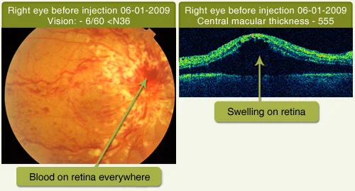
Home Central Retinal Vein Occlusion Treated With Intravitreal Injection This case is of a 54 years old Male patient. He complained Diminished of Vision for distance in Right Eye since 8 days. His vision was 6/60 Fundus examination of Right eye showed Blood on retina everywhere (Central retinal vein occlusion). He was given 2 Intravitreal Avastin injections in right eye. After Intravitreal injection his vision improved with counting finger 3mt N24 & Optical coherence tomography (OCT) done which was showed 167 microns macular thickness
Central Serous Retinopathy
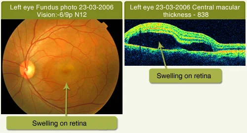
Home Central Serous Retinopathy This case is of a 33 year old Male patient He complained of Mild Blurring of Vision in Left Eye. His vision was 6/9p N12. Fundus examination of Left eye showed Swelling on Retina (Central Serous Retinopathy) Fundus fluorescence angiography (FFA) was done which showed Central Serous Retinopathy. Optical coherence tomography (OCT) showed 838 microns macular thickness. After 6 months his vision improved to 6/5 N6 without any treatment. Optical coherence tomography showed 194 microns macular thickness.
Diabetic Retinopathy Treated With Laser Treatment
Home Diabetic Retinopathy Treated With Laser Treatment This case is of a 50 years Female He complained of Mild Blurring of Vision in Left Eye. Was seen in August 2007 with swelling on the macula (Clinically Significant Macular Edema) Vision was 6/18p N10 in both eyes. She had poorly controlled diabetes with hyperlipidemia (High fat level in the blood). She has undergone (focal & grid) lasers in August 2007 for both eyes after diagnostic tests (Fundus Fluoresceine Angiography, Optical Coherence Tomography). On last examination on 01-05-2009 she has 6/12 N10 vision in right eye & 6/6 N6 vision in left eye with markedly improved fundus & her metabolic disease (Control in sugar level) is under good control. This case highlights the usefulness of laser & metabolic control in the era of Anti VEGF drugs like Avastin, Lucentis, and Macugen injections.
Diabetic Retinopathy Treated With Surgery
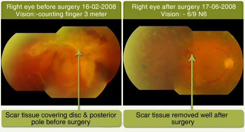
Home Diabetic Retinopathy Treated With Surgery This case is of a 58 years Female patient. Diabetes of 4 years duration not under control. She complained of Blurred Vision since 10 days Right eye more than Left eye. She underwent Vitreous Surgery in right eye and has recovered well.
Diabetic Retinopathy Treated Without Treatment
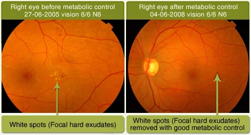
Home Diabetic Retinopathy Treated Without Treatment I would like to share with you this case where Good metabolic control ie. Control in sugar level help reverse disease process This is 37 yr. young lady. History of diabetic since age of 13 years. Her visual acuity was 6/6 N6 in 2005 (it is normal vision). The focal hard exudates (white & yellowish spots) have disappeared with good metabolic control & her vision is also maintained. Recently her (HBA1C – 6.4, Creatinine – 0.9, Lipid – Normal) diabetes and other tests were normal which means good diabetic control is required for the perfection of retina.
Endophthalmitis (Infection) Treated With Surgery
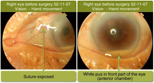
Home Endophthalmitis (Infection) Treated With Surgery This is a case of 81 years old gentleman. Had history of cataract with IOL implant surgery done elsewhere 3 years back. He had difficult cataract surgery & sutures were taken for intraocular implant insertion. After 2 years he complained of defective vision & had pus in the eye. The infection could have been due to exposed suture. He underwent Vitreous Surgery & was treated with appropriate medication & has recovered well.
Macular Hole
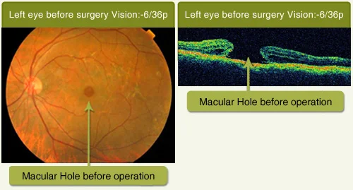
Home Macular Hole This case is of a 68 year old Female. Complained of Gradual Progressive Diminishing of Vision since a year. Her vision was counting finger 3mt, N12 improving with pinhole to 6/36part. Fundus examination Showed Full Thickness Macular Hole. Optical coherence tomography (OCT) confirmed Full thickness macular hole. She underwent Macular Hole Surgery where vitrectomy is done. Post operatively her vision improved to 6/18p N8p.
Branch Retinal Vein Occlusion (Without Any Treatment)
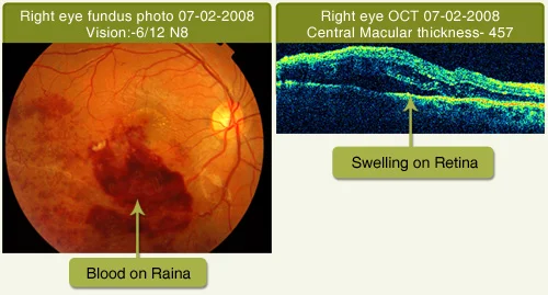
Home Branch Retinal Vein Occlusion (Without Any Treatment) This case is of a 23 years old Male patient. He complained of blurring of vision in upper left part of field of vision with right eye since 1 month. His vision was 6/12 N8 in right eye. Fundus examination of right eye showed Blood on retina with Swelling of retina (Branch Retinal Vein Occlusion with Macular edema) Optical coherence tomography (OCT) was done which showed 457 microns macular thickness. After 9 months his vision improved to 6/6 N6 without any eye treatment. Optical coherence tomography showed 194 microns macular thickness. He was given Tab. Homocheck 1 twice a day as he had hyper homocystenimia.
Macular Pucker
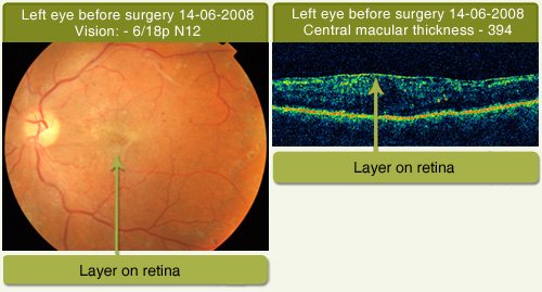
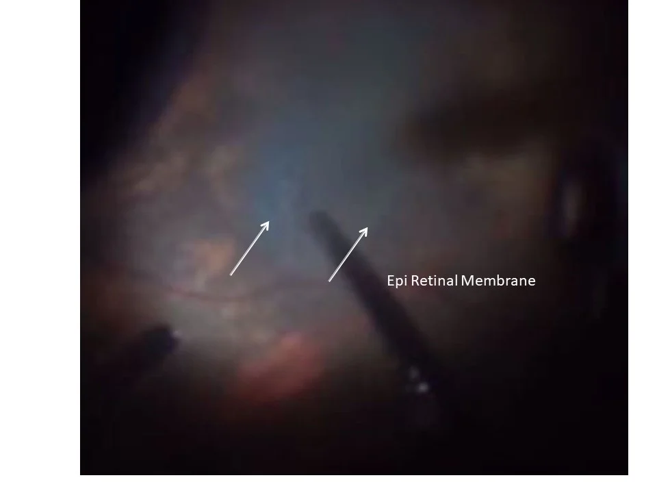
Home Macular Pucker This case is of a 67 years old Male patient. He complained of black spot & crooked vision in left eye since 15 days. His vision was 6/18p N12 in left eye. Fundus examination of Left eye showed Layer on Retina (Macular pucker) Diabetes of 30 years duration. Optical coherence tomography (OCT) done which was showed 394 microns macular thickness . He underwent Vitreous surgery for macular pucker in left eye and has recovered well with 6/9 N6 vision.
Yag Laser For After Cataract In Myopic Patients
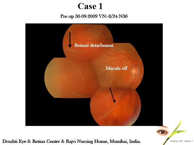
Home Yag Laser For After Cataract In Myopic Patients I would like to share the following thoughts. Recently, I attended a workshop wherein there was a discussion about the strategy to be followed for after cataract in patients with previous high myopia (Prior to cataract surgery) and it was debated whether to do Yag laser or polishing of posterior capsule. Probably no consensus was reached. I strongly feel that yag laser can be done in these patients after carefully screening the retinal periphery and treating preexisting retinal tear or lattice degeneration if any and if possible by laser treatment. If not possible, one should look at retina post laser periodically and treat if need be. I feel the results of retinal detachment surgery are quite predictable and successful in majority of patients if treated in time and treated well. The key is picking them up and fixing them at the earliest. The incidence of retinal detachment is not markedly more after yag laser. The demerit of polishing is that it requires repeated treatment and may be difficult to perform and is an intraocular surgery with all its risks.I would like to share 2 patients of acute retinal detachment following yag capsulotomy who have been successfully buckled and vision has been restored (treated in last 4 months). Case 1: He has multifocal IOL and developed retinal detachment post yag. He underwent scleral buckling and has improved to 6/12, N6 vision post op with attached retina. Case 2: He had 6/9, N6 vision, he is one eyed and developed detachment (post yag laser) temporally as seen on fundus photo and he underwent scleral buckling with core vitrectomy and has maintained 6/6, N6 vision with attached retina. I have enclosed both pre and post op fundus photo montage These patients are operated recently in last 4 months or so. I analyzed my results of scleral buckling patients last year and this year and found out interesting observations as follows. Year 2009:54 % (20/37) patients had 6/18 or better vision; & 33% (12/37) had 6/9,6/6 vision post scleral buckling operation. Year 2008 57% (12/21) patients had 6/18 or better vision; and 33% (7/21) had 6/9,6/6 vision post scleral buckling operation. These figures are small but they do point that result of early surgery in retinal detachment yield better results. I believe if patient can follow up properly for screening, yag laser does not create that big problem and if patient develops detachement and is treated in time results are very satisfying. The caution is it may not be overdone.