
Diabetic Maculopathy With Taut Posterior Hyaloid
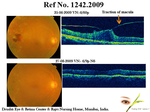
Home Diabetic Maculopathy With Taut Posterior Hyaloid 55 years old diabetic gentleman was treated with multiple lasers elsewhere for PDR. He complained of diminishing vision since 2 months his vision was 6/60p & was diagnosed by primary retinal surgeon as having cataract & macular edema & he was advised eye drops & surgery. He came to me for 2nd opinion & OCT was instrumental in correct diagnosis of vitreo macular traction & he underwent vitrectomy & recovered good vision with 6/9 N6 This case highlights the importance of OCT in all macular problems even though clinically it may not seem necessary.
Macular Pucker Case2
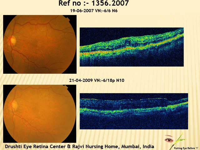
Home Macular Pucker Case2 60 years old lady presented with routine eye check up on 19-06-2007 with visual acuity 6/6 N6 in left eye & fine idiopathic cellophane maculopathy in left eye confirmed on OCT. She was asked to conserve. She came on 21-04-2009 with distorted vision in left eye. Visual acuity 6/18p N10 in left eye & with definite Epi Retinal Membrane on OCT. She underwent vitrectomy with ERM removal. 2 months post op her retina looked better but visual acuity dropped to 6/24. Patient underwent subsequent cataract surgery with IOL & fundus became better & vision improved to 6/18p N10 with no ERM on OCT. This case highlights that combined vitrectomy with Phaco could be a better choice than doing two separate surgeries as visual acuity decreased after vitrectomy due to cataract. Patient still complaint of distorted vision though anatomy has been restored well. Illustrating the need of early surgery in such type of cases with predictable results.
Lattice Degeneration
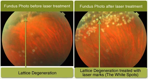
Home Recurrent Retinal Detachment This is a case of 35 year old Female Came to me for routine eye checkup and complained of floaters seen from long time. She had a family history of Retinal Detachment (brother). Her vision was 6/6 N6 with minus number glasses (short sightedness). Fundus examination showed Lattice degeneration in both eyes. She underwent Laser treatment to the Lattice degeneration area as a treatment to prevent Retinal Detachment.
Recurrent Retinal Detachment
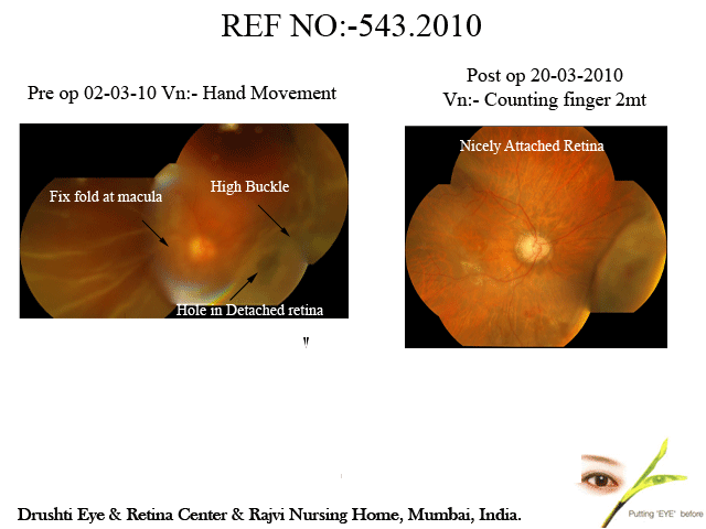
Home Recurrent Retinal Detachment This patient , 54years male came to me on 02 March 2010 complaining of Blurred vision in Right Eye and his vision was Hand movement He had undergone two retinal detachment operation in right eye in last 1 month and still had recurrent retinal detachment. He had undergone multiple operations for retinal detachment in left eye and had no vision in left eye. His two brothers had Retinal Detachment. Right eye showed Recurrent total Detachment of Retina and was operated the very same day by vitreous surgery , silicone oil injection and laser. He is under follow up and has attached retina and recovered 6/60 vision after operation. This case highlights good results with vitreous surgery even in such not so good situation.
Corneal Opacity with Detachment
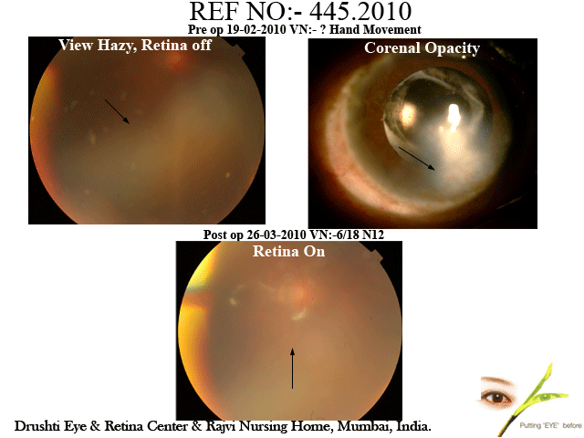
Home Corneal Opacity with Detachment This 37 year old gentleman presented with defective vision in right eye of 19 days duration in right eye on 19/2/2010, visual acuity being hand movement in right eye. He had central vascularised corneal opacities in both eyes. Left eye vision was counting finger close to face. He had total retinal detachment in right eye with early proliferative vitreoretinopathy. He underwent vitrectomy, silicone oil injection, scleral buckling and laser treatment on the same day. He has recovered 6/18, N10 vision which is very useful and has attached retina. This case highlights that with modern gadgets, such cases can also be helped by vitreous surgery.
Case Of Recurrent Retinal Detachment in One eyed patient
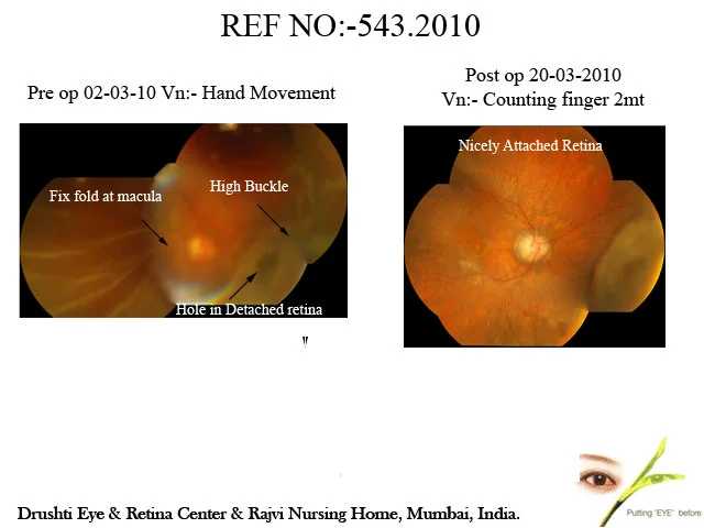
Home Case Of Recurrent Retinal Detachment in One eyed patient Th is patient , 54years male came to me on 02 March 2010 complaining of Blurred vision in Right Eye and his vision was Hand movement He had undergone two retinal detachment operation in right eye in last 1 month and still had recurrent retinal detachment. He had undergone multiple operations for retinal detachment in left eye and had no vision in left eye. His two brothers had Retinal Detachment. Right eye showed Recurrent total Detachment of Retina and was operated the very same day by vitreous surgery , silicone oil injection and laser. He is under follow up and has attached retina and recovered 6/60 vision after operation. This case highlights good results with vitreous surgery even in such not so good situation.
Case of Multiple Retinal Detachment surgries
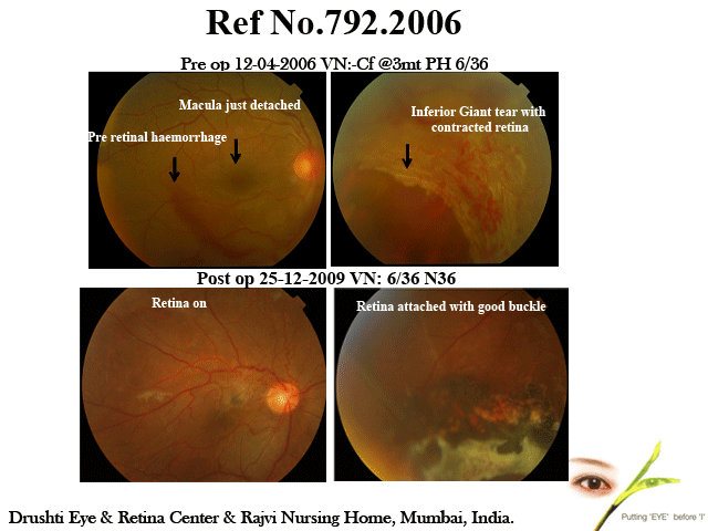
Home Case of Multiple Retinal Detachment surgries A c/o Young patient aged 17 years, With H/O cricket ball injury & defective vision of 4 days duration (on 11-04-2006) O/E AOV 6/36 IOP of 34 mm of hg Fundus : Inferior Retinal Detachment with Giant Tear & Contracted Inferior retina with macula off in right eye. On Review, Retina Attached with vision improved to 6/18 2nd surgery of silicon oil removal done on 04-10-2006 followed by Fibrin Reactio0n & Recurrent Inferior Retinal Detachment for which he underwent Re-vitrectomy with Cataract Extraction & Silicon Oil Injection on 27-11-2006. On 11/12/2009 Silicon oil was removed & secondary PCIOL placed in the sulcus. He has maintained 6/36 N24 vision with nicely attached retina with IOP under control without medication. This case beautifully illustrates good result in bad Retinal detachment with proliferative vitreo retinopathy with proper timing of surgeries.
Case of Giant Tear with Retinal Detachment
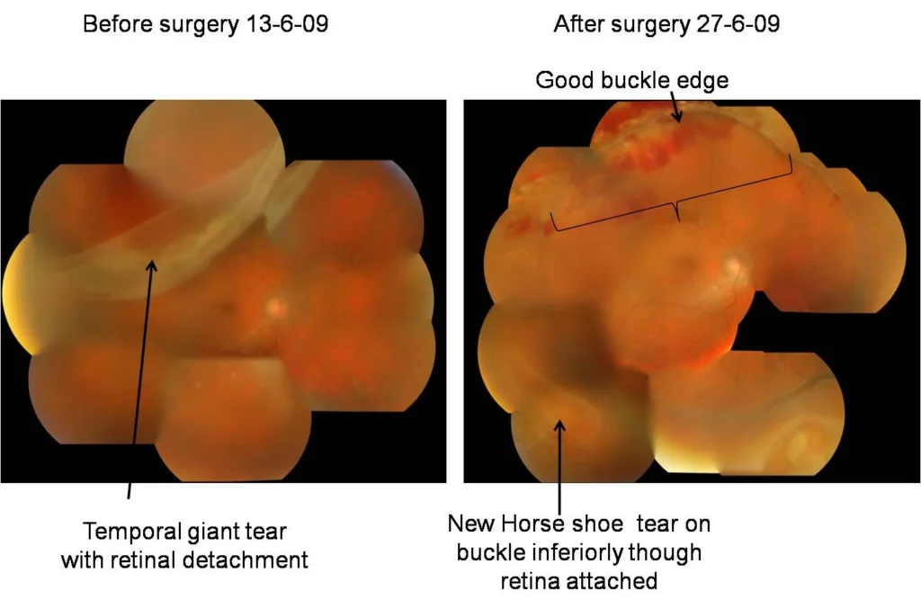
Home Case of Giant Tear with Retinal Detachment I would like to share my view on scleral buckling in present era of modern vitreoretinal surgeries.Though vitrectomies have become much advanced with better instrumentations and techniques, scleral buckling alone or in combination of vitrectomy is important in good majority of cases. This case highlights the same.This patient as seen in preop. Photograph) had 90 degree giant retinal tear in upper temporal quadrant with localized retinal detachment. He underwent vitrectomy, PFCL injection, endolaser, silicone oil injection and scleral buckling. Retina reattached very well. However he developed inferior large break in lower temporal quadrant periphery which is on buckle and hence retina did not redetach (2nd foto) and has been successfully lasered (3rd photo). Had buckle not been there, retina could have redetached needing more surgery.
Management of Complicated Retinal Detachment
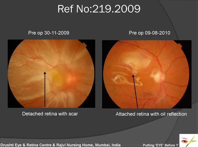
Home Management of Complicated Retinal Detachment This patient had lost vision in Rt. Eye for almost a month due to Retinal Detachment which was complicated as there were adhesions between adjacent Retinal folds. This patient underwent vitreous surgery with removal of all adhesions and reattaching Retina. It was supported by Silicone oil from inside and scleral buckle from outside. The anatomical result is excellent and patient has regained about 40% of his vision.
Traumatic Hyphema (Blood In Anterior Part Of The Eye)
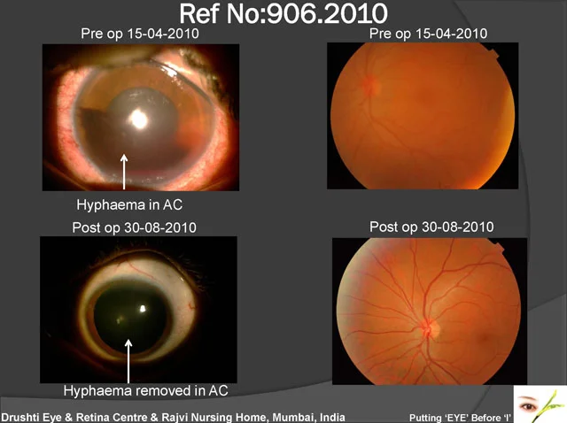
Home Traumatic Hyphema (Blood In Anterior Part Of The Eye) This 32 yr.old patient had injury to the left eye by cricket ball. He had pain & redness in the eye of 2 days duration. His vision was 6/36 and Intraocular pressure was 42 mm Hg despite all medications. He underwent washing out of blood and has recovered normal vision and pressure.