
Usefullness Of Ultrawide Field Fundus
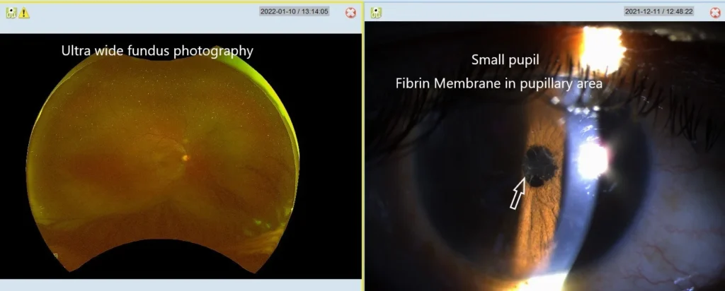
Home Usefullness Of Ultrawide Field Fundus This patient had iritis & small pupil. With normal +20D lens & indirect ophthalmoscope such panoramic view of fundus is not possible one can see fundus in such cases with +40 D lens & indirect ophthalmoscope. But ultra-wide field camera can take picture of larger areas of fundus with the small pupil also and making diagnosis more accurate.
Usefulness of Optos
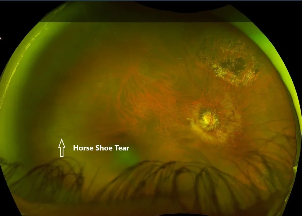
Home Usefulness of Optos This Horse shoe tear was missed clinically picked up by routine ultra wide field fundus photograph in OPTOS machine.
Usefulness Of Ultrawide Field FFA
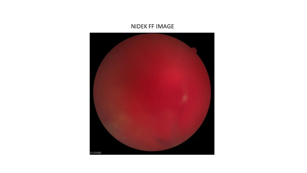
Home Usefulness Of Ultrawide Field FFA The above patient (post cataract with vitreous loss on table & PCIOL) had vitreous haemorrhage 2 months post op in right eye, fundus showed vitreous haemorrhage disc just seen, periphery visible to some extent through vitreous haemorrhage. Attached fundus photo from Nidek camera. Ultra-wide field FFA shows & confirms proliferative diabetic retinopathy with non-perfusion in periphery & leakage from NVE left eye also shows severe non proliferative diabetic retinopathy changes. This modality is useful to find the cause of vitreous haemorrhage & plan treatment.
Importance Of Identifying & Treating Cause Of CME.
Home Importance Of Identifying & Treating Cause Of CME. This 53 years old man had undergone vitrectomy with silicon oil for recurrent retinal detachment one year back & was advised ranizumab injection in left eye for cystoid macular edema elsewhere. Had 6/60 N36 vision when examined by us with cystoid macular edema, epi retinal membrane & oil emulsification. He underwent silicon oil removal with epi retinal membrane removal at our place 2 ½ months back. He has recover 6/9p N6 vision in this eye without any Anti VEGF injection & retina is attached. This illustrates importance of identifying & treating cause of CME.
Is Observation Better Than Surgery
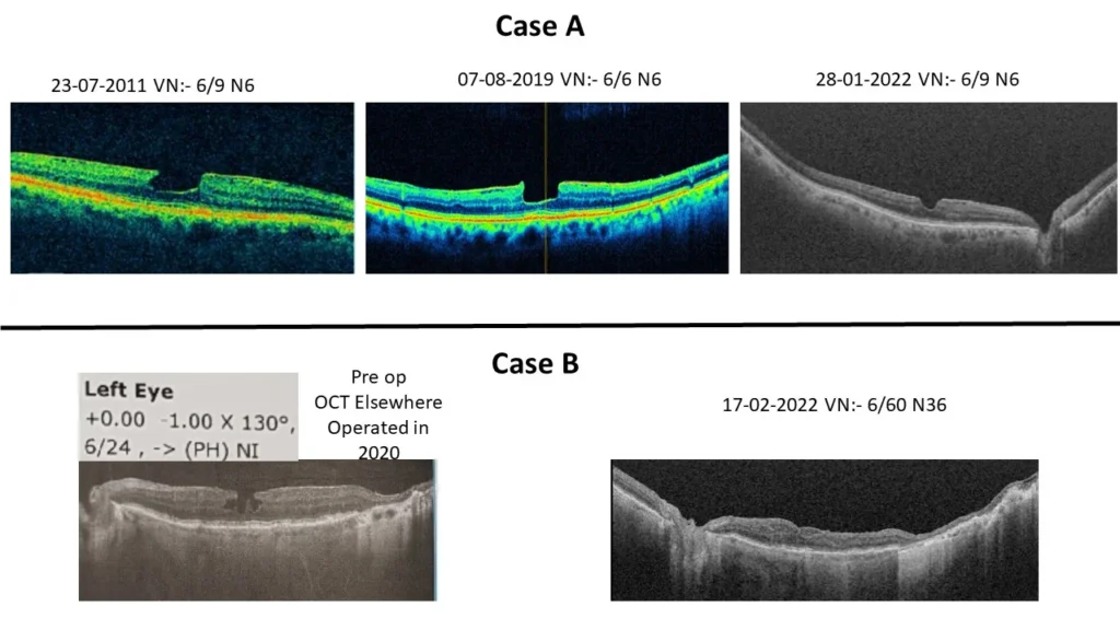
Home Is Observation Better Than Surgery Case A 84 years old gentlemen has Lamellar macular hole since last 24 years, oct done 11 years back is shown & vision is stable with 6/9 N6 without any treatment. Case B 78 years old female had undergone vitrectomy surgery for Lamellar macular hole elsewhere in 2020. Her pre-operative vision was 6/24 elsewhere & now post operatively her vision was 6/60 N36 , though anatomical result is good. The question posed is whether to operate lamellar macular hole or wait and see the natural course and decide for surgery.
Combined Retinal Detachment
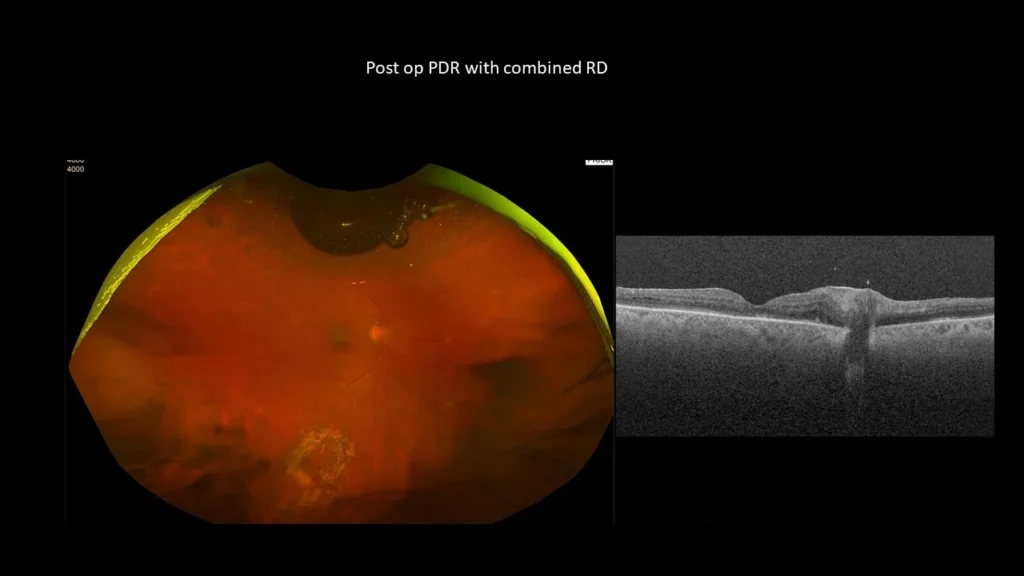
Home Combined Retinal Detachment
Montage Ultrawidefield oct A VS Optos ultrawide fundus foto
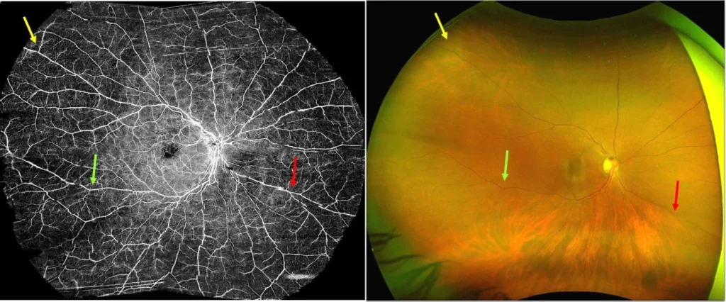
Home Montage Ultrawidefield oct A VS Optos ultrawide fundus foto Montage Ultrawidefield oct A with swept source oct shows same area as seen optos ultrawide fundus foto, arrows show same vessel marking.
Ultra wide field swept source oct A , comparing it with Optos ultra wide fundus photo
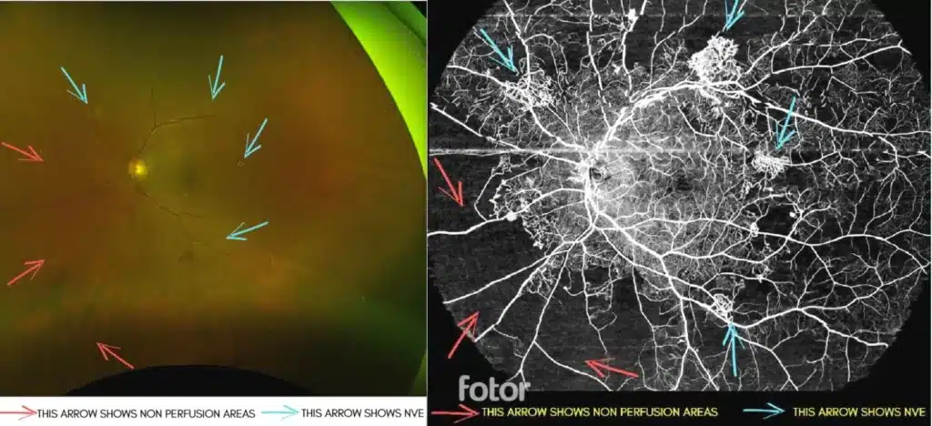
Home Ultra wide field swept source oct A , comparing it with Optos ultra wide fundus photo Would you like to share image of ultra wide field swept source oct A , comparing it with ultra wide fundus photo in a the patient with proliferative diabetic retinopathy, single picture of OCT A covers 70% of retina approximately. We are fortunate to have this technology with the grace of God. Silver stone with ultra wiidefield oct A probably makes the best imaging system today. If you enlarge fundu’s photo you will be able to see these new vessels