
Reasonably Good Despite Wet Age Related Macular Degeneration (Disciform Scar)
Home Reasonably Good Despite Wet Age Related Macular Degeneration (Disciform Scar) 71 years old gentleman with bilateral (Both eyes) Wet Age related macular degeneration. still has 6/60 visual acuity & N6 vision with (low vision aid) in right eye with early cataract This is to illustrate that though this condition is uncurable vision can be resonably good in many cases, to persue day to day activites resonably well.
Follow up Must In Retina Patient’s
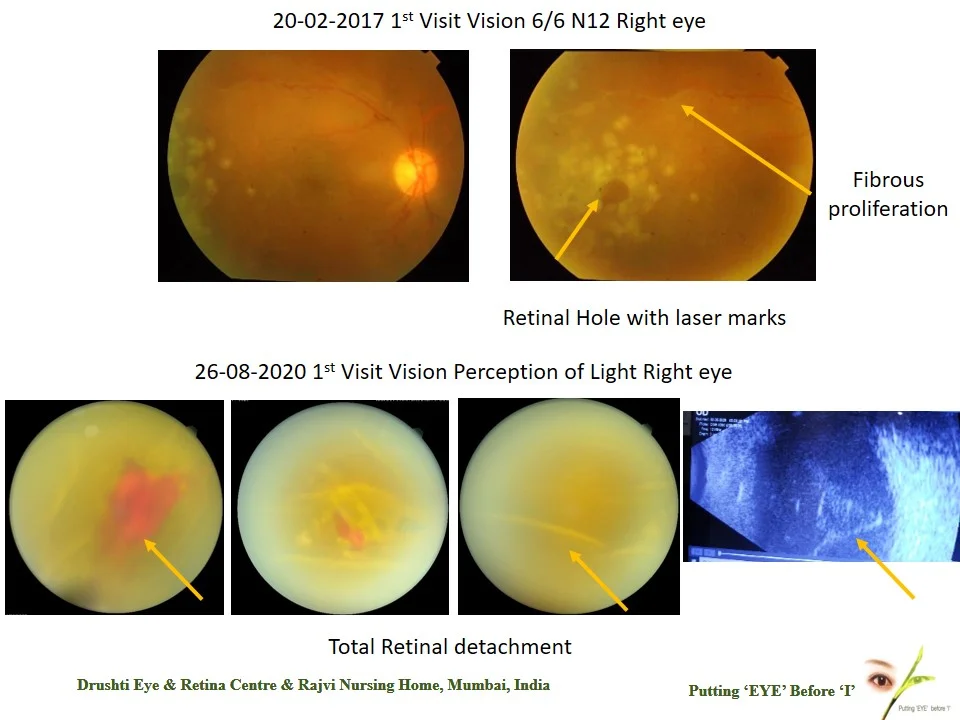
Home Follow Up Must In Retina Patient’s This 40 year old male came to me on 20-02-2017 with complaint of cobweb like thing seen in right eye since a month back His visual acuity was 6/6 N12 in both eyes Fundus showed fibrous proliferation with 3 holes & subclinical detachment ( minimal) in right eye & both eyes had healed periflebitis Same day prophylactic laser treatment was done for Right eye retinal hole with minimal retinal detachment Then he came to me on 26-08-2020 with no follow up in between On 26-08-2020 he came with complaint of blurring of vision & redness in right eye since 15 days His visual acuity was Perception of light in right eye & left eye was 6/6p Fundus showed Total detachment with Rubeosis in right eye & healed choroiditis in left eye This case is to illustrate importance of follow up in such case so that timely treatment can be given
Proliferative Diabetic Retinopathy
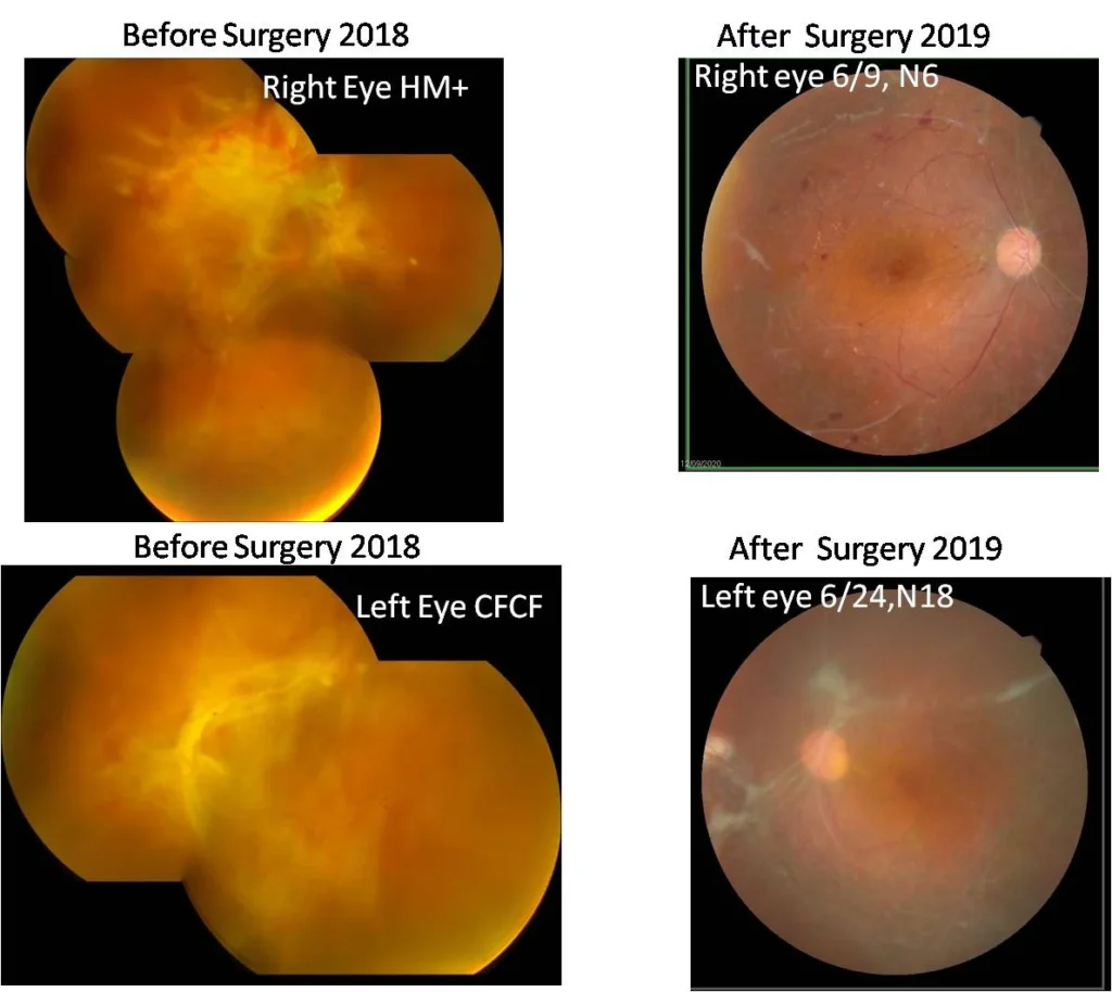
Home Proliferative Diabetic Retinopathy This 48 years old male came to me for second opinion. He was diagnosed vitreous haemorrhage elsewhere His visual acuity was right eye hand movement & left eye counting finger. He was diagnosed both eyes tractional retinal detachment and proliferative diabetic retinopathy(PDR) He was given Intravitreal Avastin injection in both eyes prior to surgery He underwent both eyes vitrectomy surgery for PDR on 07-12-2018 in right eye & 19-03-2019 in left eye respectively. Then he underwent both eyes cataract surgery in 2019 He recovered with 6/9 N6 in right eye & 6/18 N18 in left eye. Right treatment at right time can be very helpful in restoring eyesight in diabetic retinopathy cases.
Importance Of Complete Eye Examination Before Cataract Surgery
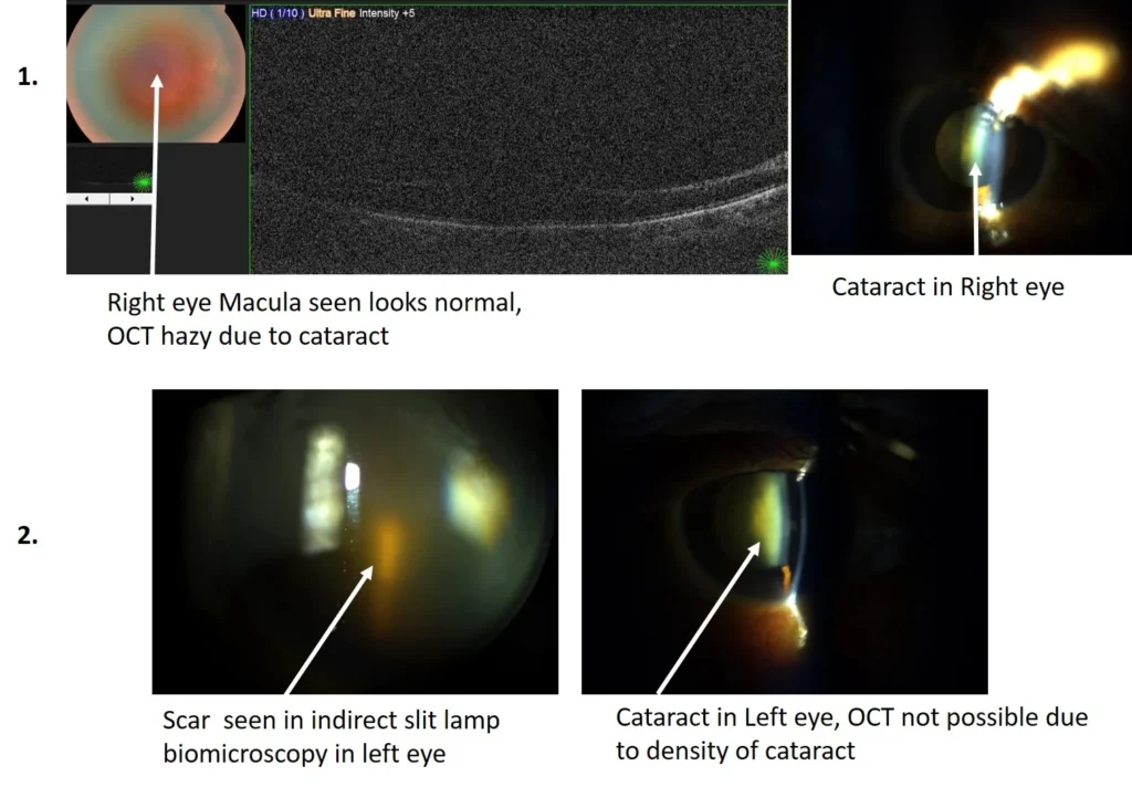
Home Importance Of Complete Eye Examination Before Cataract Surgery This 68 years old male came to for routine eye check up His visual acuity counting finger 2mt N12 in right eye & counting finger 1ft Both eyes cataract , Left eye Denser then Right eye You can see picture 1. of right eye OCT image showed normal macula but picture is hazy due to cataract and retina normal on fundus foto though view hazy. You can see picture 2. there is scar on macula which is seen in slit lamp microphotography but OCT not possible due to density of cataract. The scar on macula in left eye is visible only on indirect ophthalmoscopy & just seen on slit lamp biomicroscopy. Left eye cataract surgery will not recover good vision as there is scar on macula, no matter even if premium IOL is inserted. This case highlights importance of complete eye examination before cataract surgery to avoid unhappy patient post operatively.
Importance Of Early Removal Of Foreign Body
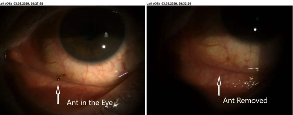
Home Importance Of Early Removal Of Foreign Body This 50 years old male came to me with complaint of irritation & foreign body sensation in left eye. He was standing in balcony & suddenly he felt something went in his eye @ 8.00pm. He called immediately & came for eye check up. There was an ant in the eye which was removed immediately & he is normal. This shows importance of early removal of foreign body
Case of Proliferative Diabetic Retinopathy
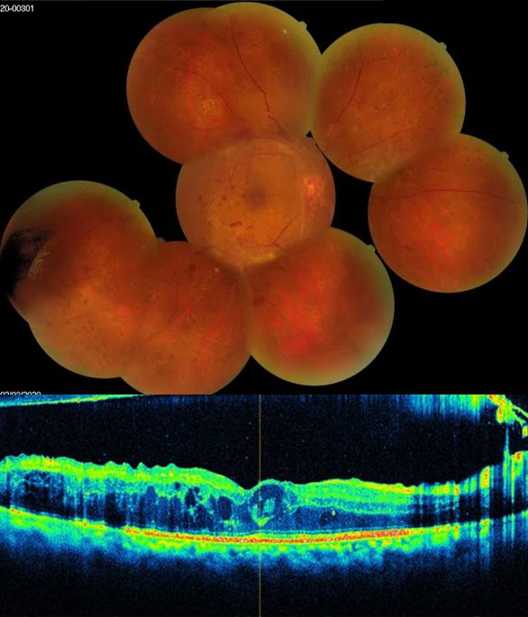
Home Case of Proliferative Diabetic Retinopathy
Role Of Pan Retinal Photocogulation In Present Era Of Anti-vegf Injection
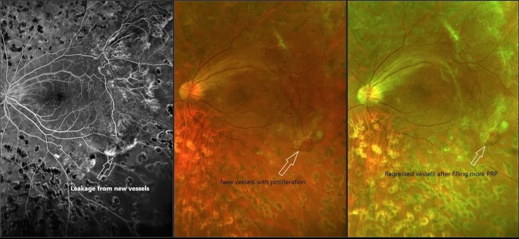
Home Role Of Pan Retinal Photocogulation In Present Era Of Anti-vegf Injection This one eyed patient with useful vision has had angry looking new vessels outside macula. He had already laser treatment to his eye Instead of giving anti-VEGF injection it was prefered to augment pan retinal photocogulation based on ultra wild field angiography. Within a month the vessels have regressed and the eye condition was stable. This shows the role of laser treatement in present era.
Yag Laser Vitreolysis
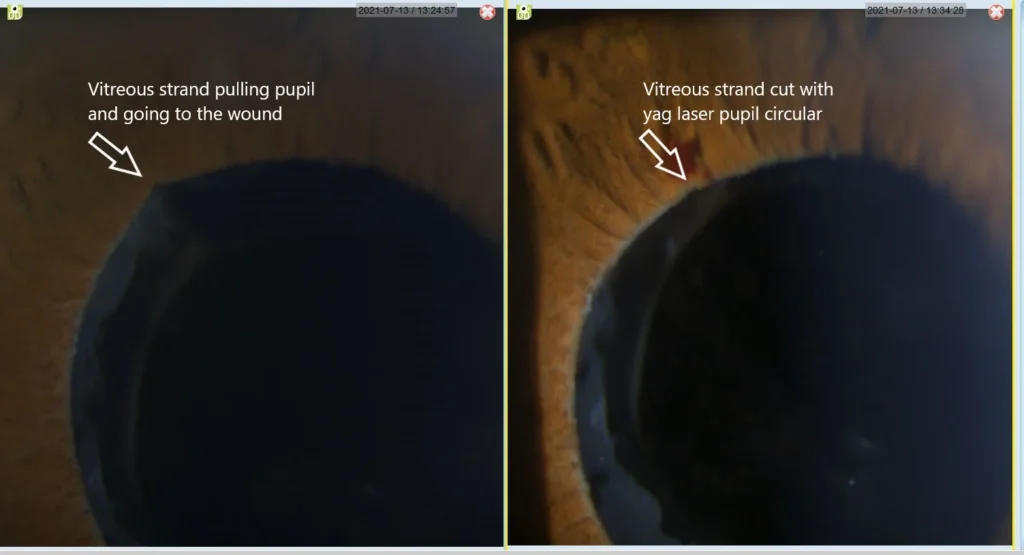
Home Yag Laser Vitreolysis This patient had posterior capsulat rupture during phaco emulsification it was managed by good vitrectomy and posterior chamber sulcus fixation IOL. Post operatively there was a residual vitreous strand pulling the iris at 10 O’clock position casusing peaking of pupil. This was successfully managed by yag laser vitreolysis. It can cause retinal detachment, decentration of IOL if untreated. It could have been treated by vitrectomy but yag laser avoided surgery.
Corneal Abrasion Role Of Fluorescein Staining
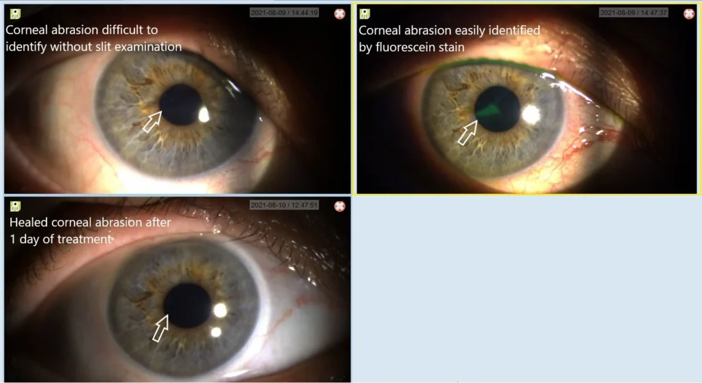
Home Corneal Abrasion Role Of Fluorescein Staining This lady had injury to right eye with hand of grand son an hour before and complained of pain. On examination she could not tolerate light and she had corneal abrasion seen on slit examination & not seen on diffuse illumination Fluorescein staining comfirmed the presence of abrasion. Her eye was padded for 1 day & she recovered. This shows importance of proper eye examination after injury especiallly if any complaint.
Usefullness Or Ultrawide Field Fundus Photo With Periferal Swept Source Oct
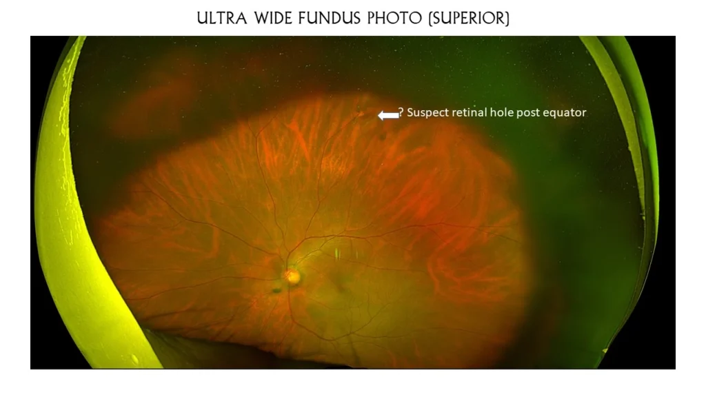
Home Usefullness Or Ultrawide Field Fundus Photo With Periferal Swept Source Oct I would like to share a case which shows the technological advancement making diagnosis accurate. This patient has symptomatic posterior vitreous detachment with operculum superiorly but no obvious retinal tear. The suspected lesion was posterior to equator so difficult to do scleral depression. Periferal OCT through lesion has confirmed the hole.