
Age Related Macular Degeneration Wet Type Treated With Inj. Avastin
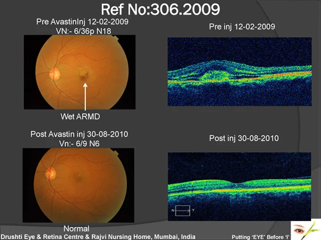
Home Age Related Macular Degeneration Wet Type Treated With Inj. Avastin This 51 year old patient had visual acuity 6/60 N18 in Left eye on 12th Feb. 2009. He was injected with Inj. Avastin in the Vitreous cavity in Feb., July, & September 2009. He is maintaining normal vision 1 & 1/2 year after treatment.
Posterior Sub Capsular Cataract With Cystoid Macular Edema
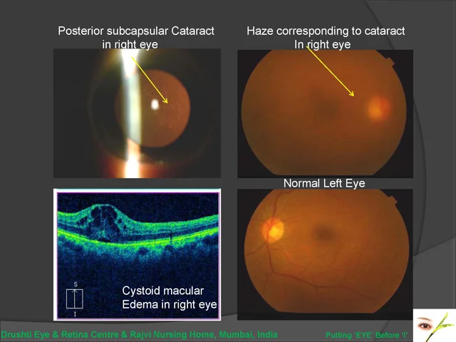
Home Posterior Sub Capsular Cataract With Cystoid Macular Edema 68 yr old female came with decreased vision in right eye, and a past history of uveitis. Vision in Right eye was 6/24p, N18, Left Eye was 6/6. Slit lamp examination showed posterior subcapsular cataract in both eyes, R > L. Fundus examination was hazy in RE due to cataract. An OCT was done to rule out other causes for reduced vision, and it showed cystoid macular edema in Right Eye. The macular edema was initially treated, following which patient will undergo cataract surgery. This case highlights the role of OCT in evaluation of patients with reduced vision. If cataract surgery had been done without diagnosing macular edema, the vision would not have improved even after surgery.
Advanced Diabetic Eye Disease Operated 20 Yrs Back
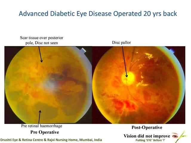
Home Advanced Diabetic Eye Disease Operated 20 Yrs Back This patient with advanced proliferative diabetic retinopathy was operated for vitrectomy in 1991 It was a difficult surgery, though successful Patient’s vision did not improve due to less circulation in the eye which had affected the optic disc. The vision could have been preserved if the condition had been detected & operated early.
Advanced Diabetic Eye Disease, Young Patient With Excellent Recovery Ref No:- 99-01196
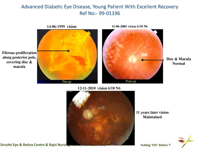
Home Advanced Diabetic Eye Disease, Young Patient With Excellent RecoveryRef No:- 99-01196 This 33 years old diabetic patient presented in 1999 with a drop in vision since 2 months. He was operated for vitrectomy in both eyes. Following a good surgery there was excellent visual & structural recovery which is maintained till today, 12 years later.
One Eyed With Advanced Diabetic Eye Disease With Good Pre Op Vision Ref No:- 06-00562
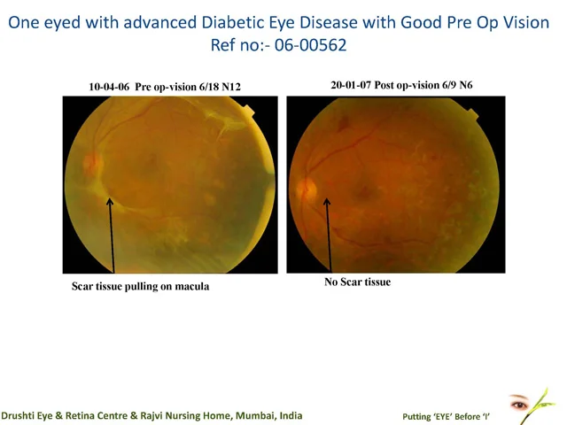
Home One Eyed With Advanced Diabetic Eye Disease With Good Pre Op VisionRef No:- 06-00562 This 48 years old one eyed diabetic patient presented with difficulty in reading since 2 months. Though pre operative vision was good 6/18 N12, there was scar tissue pulling on the macula. Post operative he recovered well & his vision improved to 6/9 N6. This stresses the need for early vitreous surgery.
Bimanual Vitreous Surgery, Quick & Effective Method
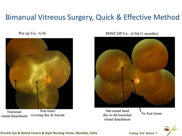
Home Bimanual Vitreous Surgery, Quick & Effective Method Routine vitreous surgery takes more than 2 hours Because of Bimanual technique, the vitreous surgery took only 50 minutes to complete.
Burnt Out Retinopathy
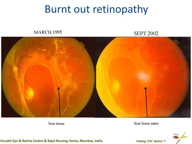
Home Burnt Out Retinopathy This 70 years female, one eyed with 6/18 vision was not offered any treatment as the proliferative diabetic eye disease was in burnt out stage & macula was not detached She maintained same vision for 8 years till last follow up.
Ref No:-06-01334 Importance Of Timely Vitreous Surgery
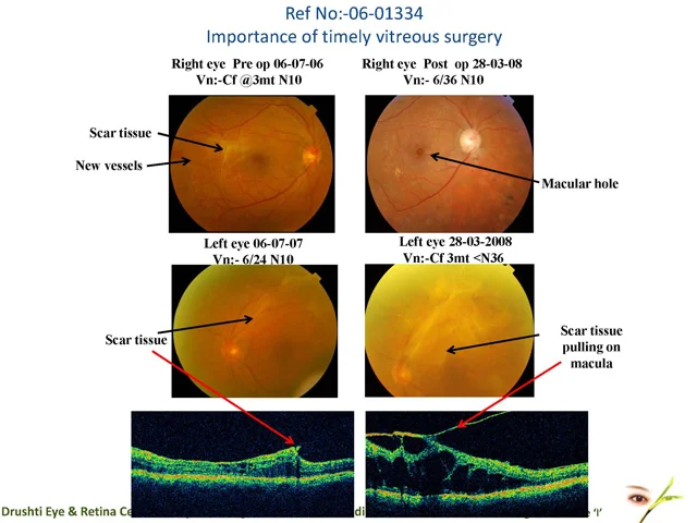
Home Role of Combination Treatment and follow up in diabetic eye diseaseRef No:-96-01291 This 60 years old male presented with proliferative diabetic retinopathy in both eyes. His vision in Right eye was Counting Finger @ 3 mt, N10, he had new vessels and fibrous tissue over posterior pole. After laser, the new vessels regressed but traction on macula increased, hence he underwent vitrectomy. Postop the vision was 6/36, due to macular hole. In the left eye, the vision was 6/24, N10. This eye was not operated and vision deteriorated to CF 3mt, < N36, in 8 months. This was due to fibrous tissue causing traction at the macula. This case emphasises the importance of timely vitreous surgery, to prevent deterioration of vision.
Role of Combination Treatment and follow up in diabetic eye diseaseRef No:-96-01291
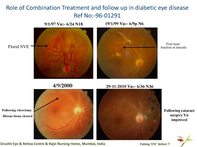
Home Role of Combination Treatment and follow up in diabetic eye diseaseRef No:-96-01291 This 34 years old patient presented with proliferative diabetic retinopathy in 1997 He had florid new vessels & fibrous tissue in the right eye. Following laser there was scarring & regression of new vessels However patient complained of distorted vision post laser which was due to traction at macula. He underwent vitrectomy in 2000. 3 years later his vision dropped due to cataract & he underwent cataract surgery. Following this combined treatment of vitrectomy & cataract surgery his vision improved to 6/36 N36 & has maintained same over 13 years.
Role Of Oct & Diabetic RetinopathyRef No:-05-01922
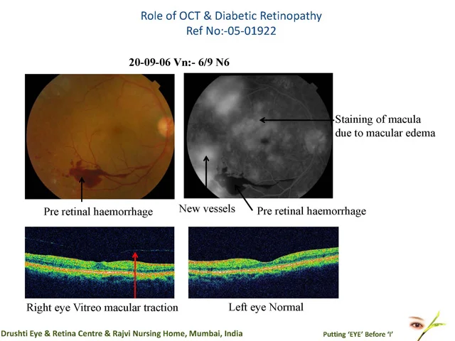
Home Role Of Oct & Diabetic RetinopathyRef No:-05-01922 This 65 years old male presented with proliferative diabetic retinopathy & 6/9 N6 vision Fluorescein angiography showed new vessels & staining of macula. One would have been tempted to give anti VEGF injection but OCT showed traction at macula which was the cause of dye leakage. Patient was observed because of good vision.