
Multiple Surgeries For Retinal Detachment
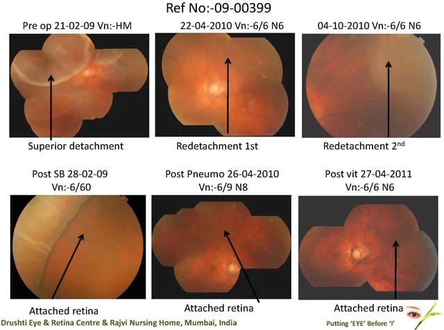
Home Role Of Oct In Evaluation Prior To Cataract Surgery 56 year old male presented on 21/2/09 with blurred vision in right eye since 3 days VA was Hand Movement and fundus evaluation revealed a superior retinal detachment He underwent scleral buckling, and at 2 months the BCVA was 6/9 with correction 14 months later, on 22/4/10 he had a redetachment superiorly and pneumoretinopexy was done The vision improved to 6/6p post pneumoretinopexy 6 months later on 4/10/10, he had a recurrent detachment of upper nasal retina, with a break at 4 oclock, though his vision was 6/6,N6 He underwent vitrectomy with C3F8 gas. BCVA improved to 6/6, which is maintained till date This case summary shows that, timely diagnosis and enthusiastic treatment can achieve excellent results even after multiple surgeries.
Role Of Oct In Evaluation Prior To Cataract Surgery
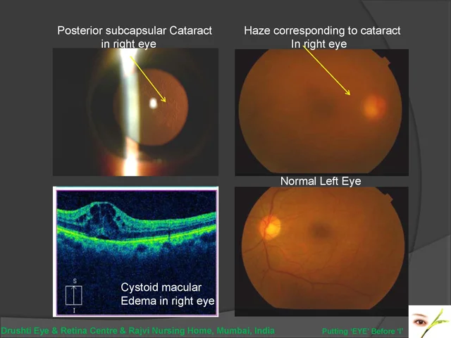
Home Role Of Oct In Evaluation Prior To Cataract Surgery 68 yr old female came with decreased vision in right eye, and a past history of uveitis. Vision in Right eye was 6/24p, N18, Left Eye was 6/6. Slit lamp examination showed posterior subcapsular cataract in both eyes, R > L. Fundus examination was hazy in RE due to cataract. An OCT was done to rule out other causes for reduced vision, and it showed cystoid macular edema in Right Eye. The macular edema was initially treated, following which patient will undergo cataract surgery. This case highlights the role of OCT in evaluation of patients with reduced vision. If cataract surgery had been done without diagnosing macular edema, the vision would not have improved even after surgery.
Enthusiastic Treatment For Post Operative Endopthalmitis
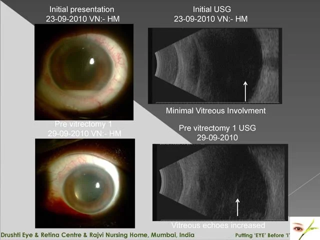
Home Enthusiastic Treatment For Post Operative Endopthalmitis Post op pseudomonas endophthalmitis presented on day 3 of cataract operation. Treated with intravenous vancomycin & ceftazidime & dexamethosone along with intravitreal antibiotics 2 injection over 5 days. Underwent first core vitrectomy on day 6 of presentation. Complete posterior vitreous detachment occurred subsequently. Underwent 2nd vitrectomy after 9 days of 1st vitrectomy. Recovered full vision 6/9 N6 within 6 weeks of treatment
Importance Of Proper Pre Op Examination
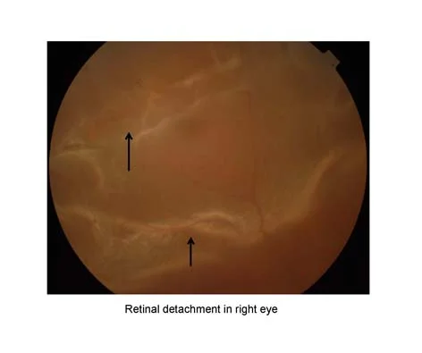
Home Importance Of Proper Pre Op Examination Patient presented with scotoma of 4 days duration in right eye & has 6/9 N6 vision with Acute superior retinal detachment What is unusual about this detachment? Patient has undergone bilateral phaco with IOL surgeries & recently yag laser capsulotomy both eyes. Patient has multiple rows of lattice degeneration in left eye which is not still detached! Patient would have had lattice degeneration in right eye also which if could have been lasered before or after cataract operation or before yag laser or after yag laser, one would have avoided this detachment Prophylactic treatment of lattice degeneration prior to cataract surgery is age old practice. If everyone learns & sees perifery of retina with indirect opthalmoscopy which is not so difficult this could definitely be avoided. Though we all are good surgeons we need to become good clinicians
Delay In Treatment Of Re-detachment Leads To Failure Or Partial Sucess
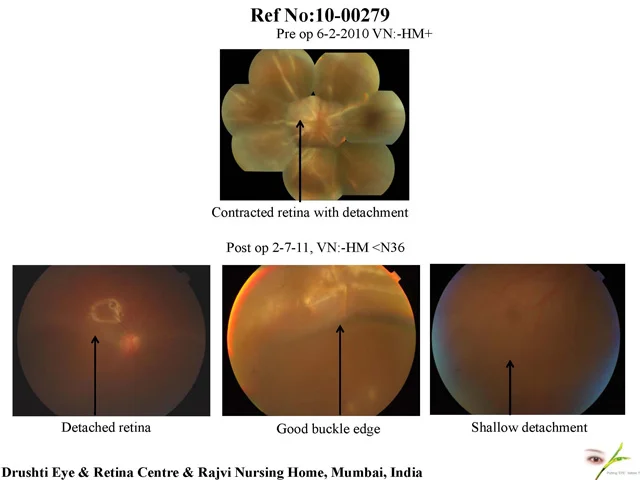
Home Delay In Treatment Of Re-detachment Leads To Failure Or Partial Sucess This 17 yr old boy presented to us after being operated for retinal detachment elsewhere, 21/2 months prior to his visit to us At presentation, his VA in RE was HM+, and fundus examination showed a retinal detachment with PVR D3 He underwent 2 revitrectomies at our place for recurrent retinal detachment and cataract extraction for mature cataract At the last follow up 2 days back, his VA in RE was HM+ This case highlights the importance of timely referral and resurgery. Due to a gap of 2 and a half months between the 1st 2 surgeries, the retina developed scar tissue and does not remain completely attached
Combined Retinal Surgery For Macular Pucker
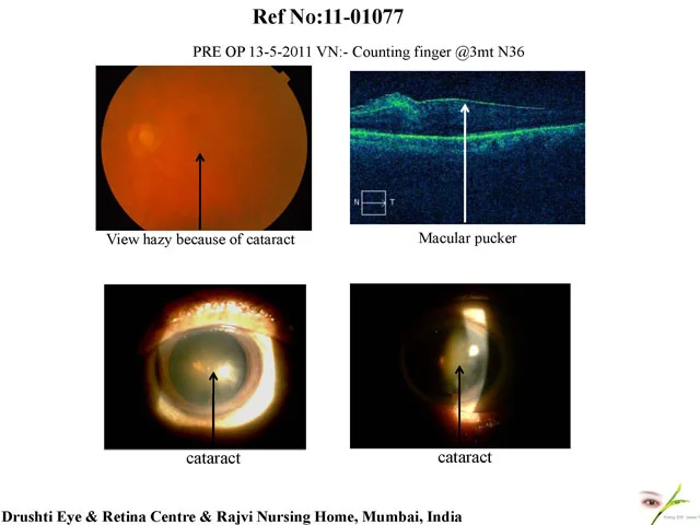
Home Combined Retinal Surgery For Macular Pucker Ref no (11-01077) 77 years old gentleman came to me on 13/5/2011 with the complaints of Left eye diminished vision, and distorted vision since 2-3 months. On examination, his left eye vision was CF-3mts;N36. Fundus examination revealed macula dull, view hazy due to cataract. He underwent OCT which showed vitreomacular traction. He underwent Phaco with IOL with Vitrectomy on 24-5-2011. Post operatively he was doing well, his left eye improved to 6/12;N6. This case highlights the importance of combined retinal and cataract surgery for macular pucker, with good postoperative results.
Macular Hole
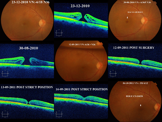
Home Macular Hole This 64 yr old female presented on 23rd Dec 2010, with a full thickness macular hole in right eye and BCVA of 6/18, N36. She was advised vitrectomy for the same, however she refused surgery at that time. She came for follow up 8 months later in Aug 2011. At that time the BCVA in RE dropped to 6/36p, N36 and hole had increased in size. On 6th September 2011 she underwent vitrectomy with sf6 gas injection and was advised prone position for 3 days as per routine postoperative instructions. However when she came for follow up 6 days later on 12th September, the hole had opposed, but was not completely closed. She was advised strict position for 22 hours, and the next day the hole had opposed much more. This position was continued for 3 more days, and on 16th September 2011, the hole was completely closed anatomically. At her last follow up visit on 4th october 2011, the BCVA in right eye had improved to 6/12, N18. This case highlights the importance of strict position, post vitreoretinal surgeries. In most patients, macular hole closes in 3 days, however in this patient it took longer probably because of the chronicity of the condition. However maintaining strict prone position helped in closure of the full thickness macular hole.
Toxoplasma Retinitis Ref No:-11-02103
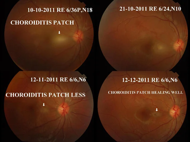
Home Toxoplasma Retinitis Ref No:-11-02103 11 yr old female, presented to us with reduced vision and seeing black spot in front of the right eye since 3-4 days. Examination showed visual acuity of 6/36, N18 in the right eye and 6/6, N6 in the left eye. Slit lamp examination and IOP were normal in both eyes. Fundus examination showed vitreous haze in right eye with an active choroiditis patch at the macula, giving a ‘headlight in fog’ appearance. She was diagnosed as toxoplasma retinitis, even though blood investigations showed normal counts, and toxoplasma titres (IgG & IgM) too were negative. She was started on T. Clindamycin 150mg BD, with tapering dose of oral steroids T. Wysolone few days later. She received 20 mg of oral wysolone for 1 week, followed by 10mg for 2 weeks, and 5 mg for 1 month. T clindamycin was continued for 2 months. Her vision in right eye improved to 6/9, N6 within 2 weeks, and she regained final vision of 6/6, N6 within a months time. The choroidits patch healed well. This case illustrate typical toxoplasma retinitis presentation
A Case Of One Eyed Proliferative Diabetic Retinopathy
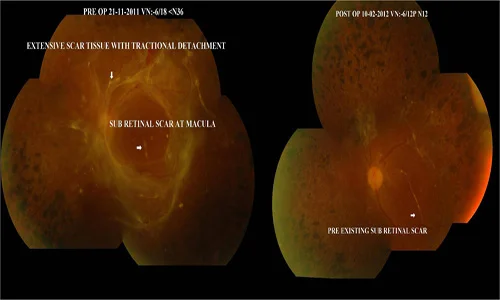
Home A Case Of One Eyed Proliferative Diabetic Retinopathy 39 year old male, known case of diabetic and hypertension on treatment, presented with reduced vision in left eye. The right eye had been operated for retinal detachment elsewhere, and he had received 3 sittings of PRP laser in both eyes. On examination, his vision in right eye was PL+, and left eye was 6/18, N36. The IOP was normal in both eyes. Fundus in right eye showed total retinal detachment, which was inoperable. The left eye fundus showed fibrous proliferation and subretinal band with tractional detachment involving the macula. He underwent bimanual 23 G vitrectomy with gas insertion. 1 month postop, the BCVA in left eye improved to 6/12p,N18 . Fundus showed disc pallor, with subretinal scar at macula, but retina was well attached. The total surgery duration was 45mins because of Bimanual 23G vitrectomy This case illustrates usefulness of timely proper vitrectomy. Patient lost the other eye with similar situation elsewhere.
Eale’s Disease Treated With Surgery
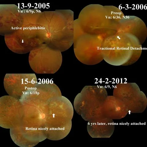
Home Eale’s Disease Treated With Surgery This 23 yr old male presented in 2005 with inferotemporal periphlebitis, and was diagnosed as Eales disease. He was started on oral steroids and AKT, however disease process remained active and 3 months later was started on immunosuppressive treatment – T.azathioprine He further worsened to develop florid NVD and tractional detachment, and underwent PRP laser followed by vitrectomy with gas insertion for the same on 17/3/06. 6 years later on follow up on 24/2/12, his visual acuity is 6/6, N6 in right eye, retina is well attached and no new lesions seen. This case illustrates usefulness of vitreous surgery in a case of vasculitis.