
Eale’s Disease Treated With Anti-koch’s
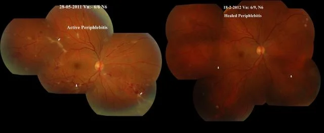
Home Eale’s Disease Treated With Anti-koch’s 23 yr old male, presented with complaints of black spots seen in front of right eye since 2 days. Past h/o TB at age of 5 yrs. VA both eyes 6/6, N6. Normal IOP. Fundus right eye showed active periphlebitis, left eye healed periphlebitis. FFA done to confirm the diagnosis of Eales disease. Investigations done – showed positive mantoux test, positive HRCT chest, and negative Se. ACE levels. He was started on oral steroids t.wysolone 60mg and full course of AKT. Steroids were gradually tapered over 4 months, and he took AKT for six and a half months. At last follow up on 18/2/2012, his vision was 6/6p, N6 in both eyes. Fundus in right eye was normal, left eye had only occasional haemorrhages. This young male patient with Eales disease, responded well to combination of oral steroids and AKT, and vascultis healed well.
Good Outcome After Multiple Surgeries For Retinal Detachment Ref No – 11-00604
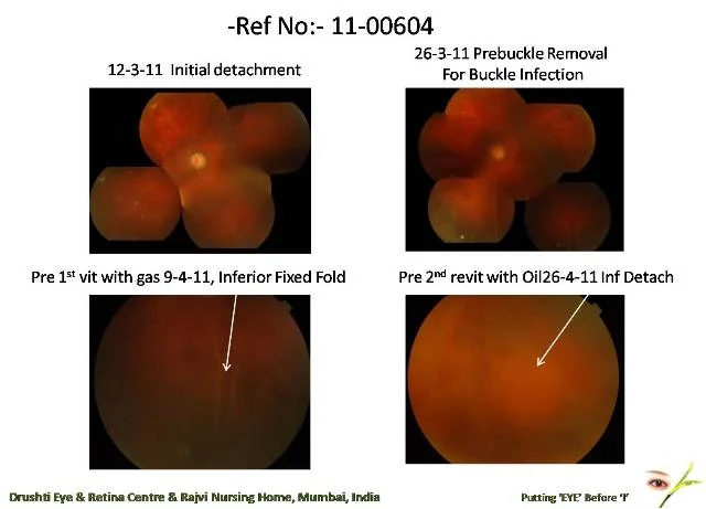
Home Good Outcome After Multiple Surgeries For Retinal Detachment Ref No – 11-00604 This 47 year old male, presented on 12-3-2011, with reduced vision in left eye since 1 ½ months. His BCVA was HM in right eye and CF 1mt in left eye. He was pseudophakic in both eyes, and left eye fundus showed lower temporal detachment. He underwent scleral buckling on the same day, however had buckle infection 10 days later with inferior Retinal Detachment. Buckle was removed, but he had inferior fixed folds, and vitrectomy with gas was done on 11-4-11. recurrent inferior detachment occurred 2 weeks later, and he was operated for vitrectomy with oil on 2-5-11. The retina remained well attached thereafter. He had a Posterior Capsular Opacity with emulsified oil on lens, so underwent posterior capsular polishing on 1-11-11, following which he had oil in AC, so oil removal was done on 6-2-12. However post oil removal, he had temporal detachment for which pnemoretinopexy on 17-2-12 and cryopexy on 24-2-12. On 28-2-12, revitrectomy with C3F8 was done. Post 3 vitrectomy, retina has been well attached for past 5 months, and vision maintained at 6/18p, N12. This case illustrates that inspite of multiple surgeries, 9 procedures done in last 1 year, retina is well attached and good vision is maintained.
Vitrectomy For Recurrent Retinal Detachment With Pvr D2 Ref. No 08-00537
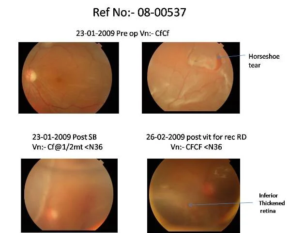
Home Vitrectomy For Recurrent Retinal Detachment With Pvr D2 Ref. No 08-00537 This 65 year old male presented with drop in vision in left eye since 15-20 days. He was psudophakic, the BCVA in left eye was CFCF, and retina showed upper temporal horse shoe tear with retinal detachment involving macula. He underwent scleral buckling on 23-1-09. The retina was attached for a month, during which there was a vitreous hemorrhage inferiorly. On 21-2-09 he developed recurrent inferior RD and underwent vitrectomy with oil on 25-2-09. Post vit he had inferior thickened retina, which led to inferior recuurent detachment. On 6-4-09, the BCVA was CFCF, and there was RD with PVR D2. On 7-4-09, he was operated for vitrectomy with 180 degree inferior retinectomy, with silicon oil and endolaser. Post op, the retina remained well attached. Silicon oil was removed on 15-12-11, and on the last follow up on 23-1-12, the BCVA in left eye was 6/36, N18, with attached retina.
Revitrectomy For Recurrent Retinal Detachment With Pvr, Following Slipped Edge Of Giant Retinal Tear Ref No:- 11-02496
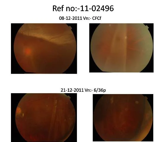
Home Revitrectomy For Recurrent Retinal Detachment With Pvr, Following Slipped Edge Of Giant Retinal Tear Ref No:- 11-02496 This 22 year old male presented on 8-12-11, with sudden drop in vision in left eye since 2 days. BCVA was CFCF, and fundus showed temporal retinal detachment with giant retinal tear. He underwent vitrectomy with silicon oil and buckle on 9-12-11. Post op, it was seen that the temporal edge of the GRT had slipped from the buckle. On further follow up, the retina remained attached, but the slipped edge started getting lifted up. On 30-12-11, he underwent revitrectomy with oil, the edge of the GRT was cauterised and cut, and retina was lasered. Post op, retina was well attached and there was no slippage. However, he subsequently developed PVR, which led to tractional retinal detachment nasally. On further follow up the detachment persisted, and on 6-7-12 he underwent 3rd vitrectomy for recurrent RD with PVR, with lensectomy. On last follow up on 6-8-12, BCVA was CFCF, but retina was well attached.
A Patient Of Macular Hole With Retinal Detachment Who Improved After Multiple Vitrectomies Ref No:-10-01527
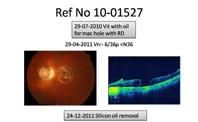
Home A Patient Of Macular Hole With Retinal Detachment Who Improved After Multiple Vitrectomies Ref No:-10-01527 This 52 year old male, presented on 20-7-10, with sudden painless drop in vision in left eye since 20 days, and black patch seen in centre of eye. The BCVA in LE was counting finger 1 ft, < N36. There was PCIOL in situ, and fundus showed macular hole with retinal detachment. He underwent vitrectomy with silicon oil for the same on 29-7-10. Post op, BCVA improved to 6/36p, < N36 in left eye. On 24-12-11, silicon oil removal was done. On 5-5-12, his vision in left eye dropped to HM. Fundus showed that macular hole was open, and there was inferior detachment with early PVR. On the same day he underwent revitrectomy for recurrent RD post SOR. However on 22-5-12, it was seen that macular was again open, and inferior fluid was present, which persisted and did not get absorbed. On 6-7-12, he underwent 3rd vitrectomy with oil for recurrent RD. The was a fine epiretinal membrane over macula, which was removed during surgery. Following 3rd vitrectomy, the macular hole remained closed. On the last follow up on 17-8-12, the vision in left eye was CF 1 mt, and macular hole was closed. There was however shallow inferior retinal off, maybe due to PVR changes.
Vitrectomy for recurrent retinal detachment post scleral buckling Ref no.11-01752
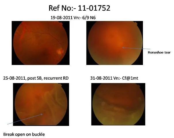
Home Vitrectomy for recurrent retinal detachment post scleral buckling Ref no.11-01752 This 40 year old female presented on 18-8-11 with sudden drop in vision in right eye since 2 days. BCVA was 6/9, N6 and fundus showed acute inferior retinal detachment, with HST at 6 Oclock. She was operated for scleral buckling on 19-8-11. Post op there was inferior fluid and break on the buckle was open. She was asked to maintain shirsashan position (head down) in the ward for inferior break for few days, however the retina developed recurrent detachment with folds. Vitrectomy with oil for recurrent RD was done on 30-8-11. Next day retina was attached. Silicon oil removal was done on 8-5-12. At her last follow up on 20-7-12, the BCVA in right eye was 6/24, N12p and retina had remained attached. Vision was probably less due to developing cataract.
Cataract With Parafoveal Telangiectasia ( Ref No 08-00919)
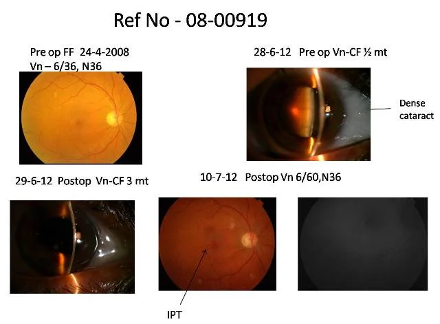
Home Cataract With Parafoveal Telangiectasia ( Ref No 08-00919) This patient initially presented in 2008, when right eye vision was 6/36, N36, and fundus showed Idiopathic Parafoveal Telangiectasia (IPT) She then came again on 14-4-12 with reduced vision in both eyes since 1-2 months. The vision in both eyes was CF ½ mt. She had significant cataract in both eyes. She underwent uneventful phaco with IOL on 28-6-12 in right eye, following which vision improved to 6/60, N36. IPT is generally a non progressive condition, hence the visual recovery was good after cataract surgery, even though it did not improve to normal.
Cataract with Parafoveal Telangiectasia ( Ref No 02-01069)
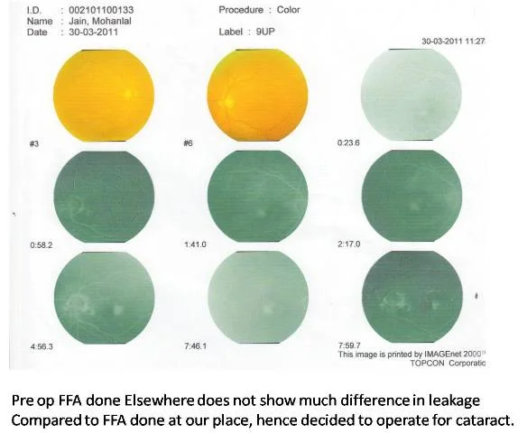
Home Cataract with Parafoveal Telangiectasia ( Ref No 02-01069) This 69 yr male, initially presented in 2002. Vision at that time was 6/9, N6 Right Eye and 6/6, N6 Left Eye. He came on 14-06-2011, with reduced vision both eyes since 6-8 months. Vision was 6/24,N36 in Right Eye and 6/18p, N12 in Left Eye. Right Eye had significant cataract. He first underwent cataract surgery -phaco with IOL in Right Eye, on 28-6-11, following which Vision improved to 6/9, N8. On 28-11-11, Left Eye was operated for cataract surgery -phaco with IOL, and Left Eye Vision improved to 6/9, N6.
Cataract With Subhyaloid Hemorrhage ( Ref No 11-00410)
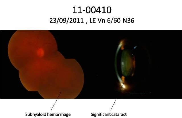
Home Cataract With Subhyaloid Hemorrhage ( Ref No 11-00410) This patient, known diabetic, presented with reduced vision in the left eye. On 23-9-11, Left eye vision was 6/60, N36. There was a significant cataract, and fundus showed subhyaloid hemorrhage. He underwent cataract surgery with phacoemulsification and Intraocular Lens Implant on 30-9-11, and after the surgery, Pan Retinal Photocoagulation (PRP) laser was done on table. Postoperatively, vision was 6/12, N18 on the next day, and at last follow up on 20-7-12, it was 6/9,N6.
Importance of OCT in preop evaluation (Ref No 09-02329)
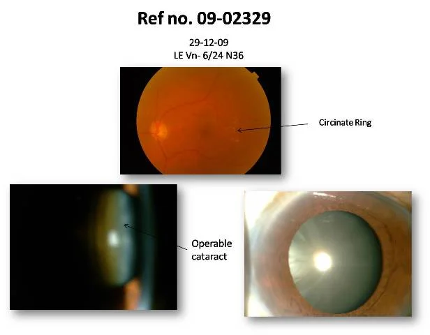
Home Importance of OCT in preop evaluation (Ref No 09-02329) This Patient came on 29-12-09 for cataract Evaluation. Vision was 6/24,N36 in left eye. There was an operable cataract in left eye, and fundus showed circinate ring of hard exudates. One would have otherwise done cataract surgery followed by intravitreal anti – VEGF injection or laser. However, a routine OCT done at our place showed Vitreo Macular traction (VMT) in the affected eye. He underwent Cataract surgery – phacoemulsification with intraocular lens implant with vitrectomy for VMT in left eye on 7-1-10. Post operatively his left eye vision on 17-3-10 was 6/6,N6