
Cataract with Diabetic Maculopathy (Ref No:- 04-01765)
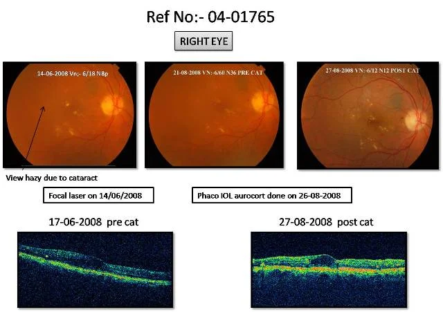
Home Cataract with Diabetic Maculopathy (Ref No:- 04-01765) This 61 yr old patient, known diabetic since 20 yrs, presented in 2008, with reduced vision in both eyes. Vision in right eye was 6/18 N8p, and in left eye was 6/9, N6. There was early cataract in both eyes, and fundus showed diabetic maculopathy. Patient initially received focal laser for the maculopathy in both eyes. Then he underwent cataract surgery in both eyes – phacoemulsification with IOL implant, with intravitreal steroid injection. Right eye was operated in Aug 2008, and Left eye operated in Feb 2010. Post op he again received focal laser in both eyes. 3 years later, at last follow up he is doing well, with final vision as 6/6, N8 in right eye and 6/6, N6 in left eye.
Cataract with Diabetic Maculopathy (Ref No:10-02468)
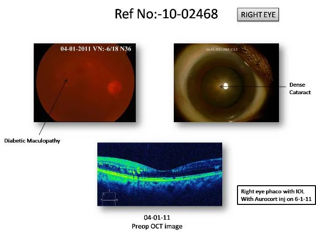
Home Cataract with Diabetic Maculopathy (Ref No:10-02468) This Patient, known diabetic, presented for cataract and retina evaluation. Vision at that time was 6/18, N36 in right eye and 6/18p, N18 in left eye. She had cataract in both eyes and fundus showed diabetic maculopathy. Right eye was initially operated for brown cataract with Phacoemulsification and IOL and steroid injection, followed by focal laser and intravitreal avastin injection, but did not improve inspite of good metabolic control. Left eye was also similarly treated, and has improved well. This case illustrates that sometimes inspite of appropriate treatment, the diabetic maculopathy does not improve.
Cystoid Macular Edema post Cataract Surgery (Ref No : 08-00866)
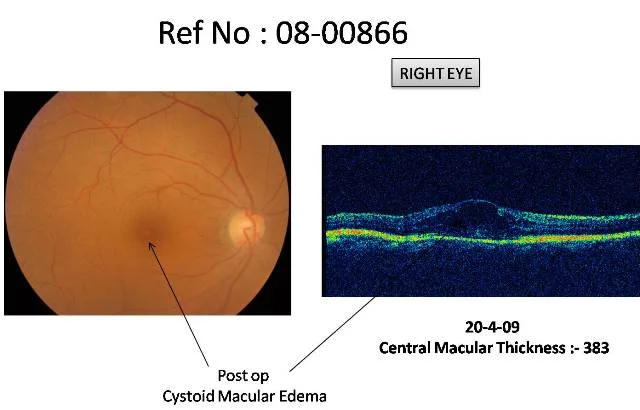
Home Cystoid Macular Edema post Cataract Surgery (Ref No : 08-00866) This 60 year old lady underwent uneventful phacoemulsification cataract surgery with foldable intraocular lens implant. At 1 month follow up, Vision in operated eye was 6/9,N6 but patient was not happy with her vision. She felt her vision was blurred and there was a film in front of her eye. OCT done postop revealed presence of Cystoid Macular Edema post surgery, which was the reason she was not happy with her vision.
Cataract Surgery Followed by Cystoid Macular Edema, treated with oral Diamox (Ref no :09-00285)
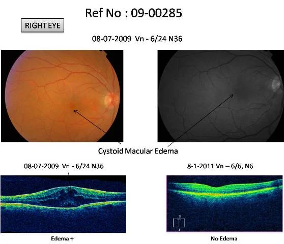
Home Cataract Surgery Followed by Cystoid Macular Edema, treated with oral Diamox (Ref no :09-00285) Patient came for cataract evaluation. Vision was 6/9p, N8p in right eye. She had cataract in right eye, and retina was normal. She underwent Right eye Cataract Surgery- Phacoemulsification with IOL implant on 23-04-09. Post operatively she was diagnosed cystoid macular edema in right eye. Her vision was 6/24,N10. She was started on eye drops and Tab Diamox orally. On last visit her vision was 6/6,N6 in right eye and edema had resolved.
Cataract Surgery followed by Cystoid Macular Edema, treated with eye drops only (Ref No:12-00995)
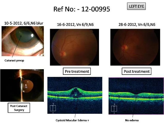
Home Cataract Surgery followed by Cystoid Macular Edema, treated with eye drops only (Ref No:12-00995) This 53 year old female came to me with complaints of left eye blur vision mainly at night. Vision was 6/9,N6 blur and slit lamp showed Posterior sub capsular cataract, Fundus was normal in left eye. She underwent Left eye Cataract Surgery – Phacoemulsification with IOL implant on 16-5-12. The surgery was uneventful. Post operatively next day vision was 6/6,N10 and fundus was normal. On 16-6-12 she complained of dull vision in left eye, vision was 6/9,N6 and fundus examination showed Cystoid Macular Edema. She was started on topical drops for the same, which resolved in 2 months. On 28-8-12, her vision in left eye returned to normal – 6/6,N6.
Cataract Surgery followed by Cystoid Macular Edema, treated with Aurocort Injection (Ref No : 04-02037)
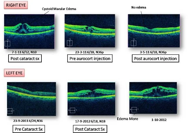
Home Cataract Surgery followed by Cystoid Macular Edema, treated with Aurocort Injection (Ref No : 04-02037) This patient underwent Right eye uneventful cataract surgery – phacoemulsification with IOL implant on 8-12-10. Preop vision was 6/18, N36p. Post op, vision was 6/18, N36 and fundus showed Cystoid Macular Edema (CME). Initially she was given medical management with T. Diamox and Megabrom eye drops. However, edema persisted for 4 months post cataract, and she was given Inj. Aurocort in RE on 26-04-11. Post aurocort, edema settled and vision improved to 6/6, N6. She underwent left eye uneventful cataract surgery – phacoemulsification with IOL implant on 20-8-12. Preop Vision was 6/60, < N36. Postop, vision improved to 6/12, N12. However a month later it dropped to 6/18,N18, and there was cystoid macular edema. This case illustrates that CME can occur in both eyes inspite of good surgery. The left eye too will probably require Aurocort injection like the right eye.
Cataract With Retinitis Pigmentosa (Ref No:08-00592)
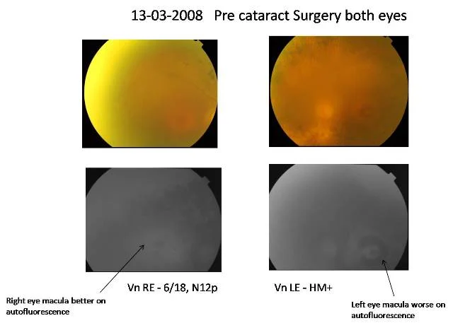
Home Cataract With Retinitis Pigmentosa (Ref No:08-00592) This 79 yr old male presented on 13-3-08 with reduced vision in both eyes since 3 months. vision in right eye was 6/18, N12p, and in left eye was Hand movement. There was significant cataract in both eyes, and fundus showed Retinitis Pigmentosa. The fundus autofluorescence showed that right eye macula was better than left eye. Considering right eye retina was better, that eye was operated first, and vision improved to 6/9,N6. Seeing the good results in right eye, the left eye was operated a year later and vision improved to Counting Fingers Close to Face. However at follow up on 29-11-10, vision in both eyes had dropped to Hand movement, due to worsening of retinopathy
Patient was advised cataract surgery elsewhere, Ethambutol toxicity was missed (Ref No 09-02009)
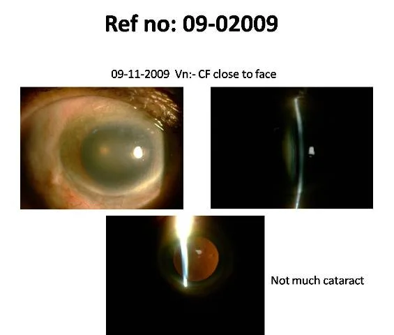
Home Patient was advised cataract surgery elsewhere, Ethambutol toxicity was missed (Ref No 09-02009) This patient had counting finger close to face vision in right eye at presentation and was advised cataract surgery, but the slitlamp photo hardly reveals any significant cataract. He had been taking ethambutol tablets and it was stopped. He improved in vision without any surgery in short time.
Postoperative Endophthalmitis (Infection) Managed With Vitrectomy With Excellent Result (Ref No:10-01965)
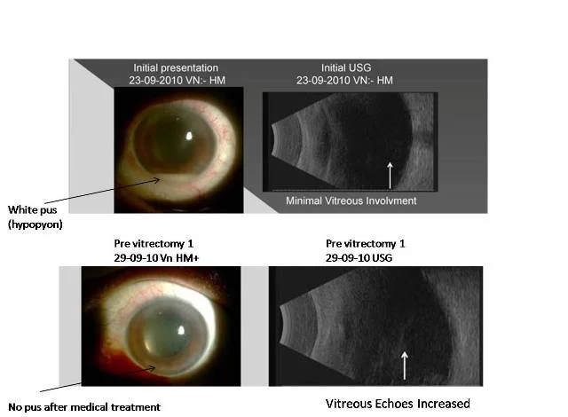
Home Postoperative Endophthalmitis (Infection) Managed With Vitrectomy With Excellent Result (Ref No:10-01965) This 56 year old male came to me on 23-09-10 with a complaint of diminished vision in left eye post cataract surgery which was done on 21-09-10. Vision in left eye was Hand movement. He had white pus (hypopyon) in the front of the eye, and fundal glow was seen. He was initially started with intravenous antibiotics. With that, the pus in front of eye had started clearing, but vitreous was not clearing so he underwent core vitrectomy on 29-09-2010. There was incomplete posterior vitreous detachment ( as also seen on Bscan done preoperatively). No vigorous attempts to induce posterior vitreous detachment were made. He underwent Revitrectomy for Endophthalmitis on 7-10-10. Post operatively he has recovered full vision to 6/6P,N6 in left eye.
Improtance Of Complete Eye Examination
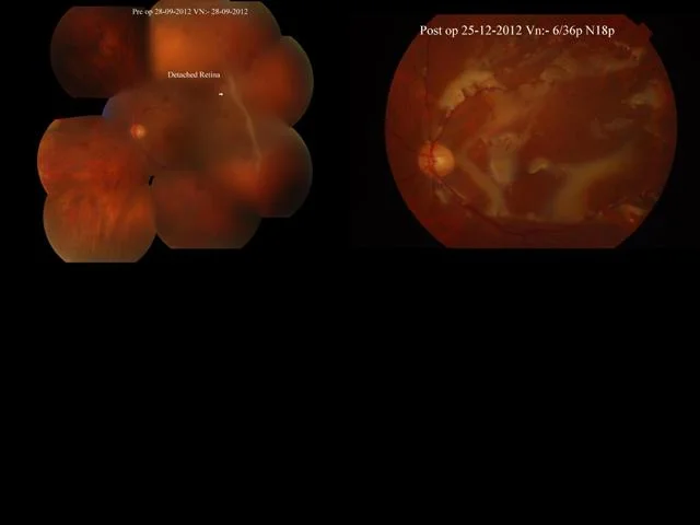
Home Improtance Of Complete Eye Examination The following is a 65 year old patient who had curtain like field defect in the right side of left eye. He had undergone some brain operation 10 years back and had undergone both eyes cataract surgery. He was advised CT scan brain in view of the field defect by the general ophthalmologist who had done his cataract surgery whereas the field defect was due to retinal detachment. He underwent vitrectomy with buckling and silicone oil injection and has improved.