
Ref No:- 12-01838 Vitrectomy for Giant Retinal Tear with Retinal Detachment
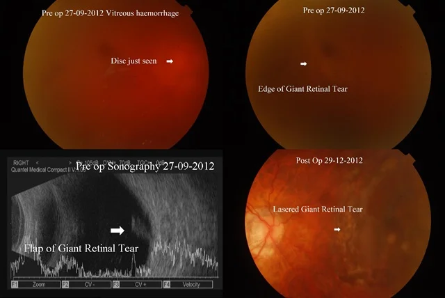
Home Ref No:- 12-01838 Vitrectomy for Giant Retinal Tear with Retinal Detachment This 58 yr old male presented to me in sept 2012 with complaints of seeing flashes and floaters in right eye since 7 days and 3 days, respectively, and a curtain like obstruction to vision since then. On examination, the vision in right eye was Counting Fingers 3mt, improving to 6/60 with pin hole. Slit lamp examination showed a cataract, and fundus showed red vitreous hemorrhage, with a suspect giant retinal tear (GRT) upper nasally. Sonograpohy showed vitreous hemorrhage, with a flap seen in upper nasal quadrant, due to the giant retinal tear. On follow up 2 days later, the hemorrhage had cleared a bit, with vision improving to 6/24p, and retina was visible, with GRT and retinal detachment seen. However laser to the tear was not possible because of the cataract. He underwent cataract extraction with vitrectomy on 1-10-12, with c3f8 gas and scleral buckling. Post op, the best corrected vision in RE improved to 6/6p, and retina has remained well attached.
Ref No:- 13-00109 Complication of Cataract Surgery
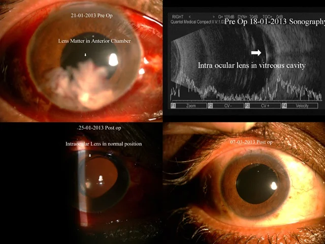
Home Ref No:- 13-00109 Complication of Cataract Surgery This patient presented on 15-01-13, with reduced vision in right eye after cataract surgery which was done elsewhere the previous day On examination, the vision was Hand movements, slit lamp showed corneal folds, with aphakia and lens matter in anterior chamber. There was a vitreous hemorrhage, and details of retina were not visible. The intraocular pressure was normal. When he came for follow up 3 days later, the findings were same, but eye pressure had gone up to 32 mm Hg. He underwent vitrectomy on 23-01-13, for retained lens matter. The vitreous hemorrhage was cleared, and intraocular lens was seen in the vitreous cavity, which was removed and placed in the normal position. Postoperatively, the vision improved to 6/6, IOL was well centered and retina was normal.
Timely Removal Of Silicon Oil
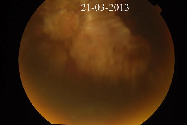
Home Timely Removal Of Silicon Oil I would like to stress the timely removal of silicon oil in patients who have undergone successful vitreo retinal surgeries. Sometimes primary retinal surgeons not remove the oil in time & patient becomes blind because of Glaucoma. the following two cases illustrate the point. Patient 1: 13-00221 This 31 yr old male patient presented to me on 4-2-13, with complains of blurred vision in right eye. He had been operated vitrectomy with oil for retinal detachment on 13-12-2010 elsewhere. He had previously undergone LASIK and multifocal IOL implants in both eyes elsewhere. The BCVA in RE was CF 3 mts and slit lamp showed multifocal implants. Fundus showed glaucomatous disc in right eye and myopic disc in left eye, with myopic macula in both eyes. He was on treatment with Combigan, Misopt and ganfort eye drops, and IOP was 13 mm Hg in RE. He underwent oil removal on 12-2-13. on 12-03-2013 vision is 6/60 N36, retina attached & patient is on Timolet eye drop 2 times a day with IOP of 20mm of hg in right eye Patient 2:- 10-02227 This 61 yrs old one eyed male presented to me on 20-11-2010 with past history of vitrectomy with silicon oil in 2008 for retinal detachment His BCVA in left eye was 6/60 N36, fundus showed myopic & glaucomatus disc & macula, retina attached, IOP 19mm of hg with Travatan eye drop once a day, Iotim eye drop 2 times a day & Dorzox eye drop 3 times a day He was advised silicon oil removal. He came again on 03-08-2012. His visual acuity dropped to counting finger @ 2mt. Funuds showed retina attached, IOP 20mm of hg with Travacom eye drop once a day, Alfagan Z eye drop 2 times a day, Dorzox eye drop times a day He was advised silicon oil removal. He came on 21-03-2013 with Hand movement vision & IOP of 34mm of hg. Patient had stopped all glaucoma treatment as he was loosing vision. In above 2 patients timely silicon oil removal could have controlled glaucoma better & saved usedfull vision. In my opinion quiet a few retinal surgeons are scared to remove silicon oil with fear of retinal redetachment. However with good previous retinal surgeries & case selections this can be minimized to a great extent by timely removal of silicon oil & we would be able to save the eye from potencially blinding glaucoma.
Secondary Glaucoma Following Retinal Surgery
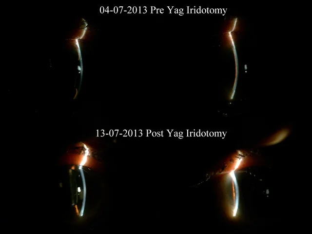
Home Secondary Glaucoma Following Retinal Surgery This 70 years old lady had undergone left eye combined cataract with vitrectomy for macular hole on 29-05-2013 elsewhere. She had pain & high pressure after about 10 days of surgery & had undergone AC reformation with gas elsewhere for shallow AC elsewhere. UBM done by Dr. Deepak Bhatt confirmed Iris bombe On presentation her vision was counting finger 1mt in left eye, flat chamber in periphery deep in centre with well-placed posterior chamber intra ocular lens. Intra Ocular Pressure was 19mm of hg in left eye with Tab. Diamox 1-1-1-1 times a day with Predfort eye drop & Atropine eye drop. Funds examination revealed gas fill 40%, closed macular hole. I did multiple yag iridotomies in periphery in left eye. 10 days after treatment anterior chamber became deep & intra ocular pressure was under controlled after stopping Tab. Diamox. This case illustrates that timely yag iridotomy can help in iris bombe post-surgical.
Role Of Optical Coherence Tomography
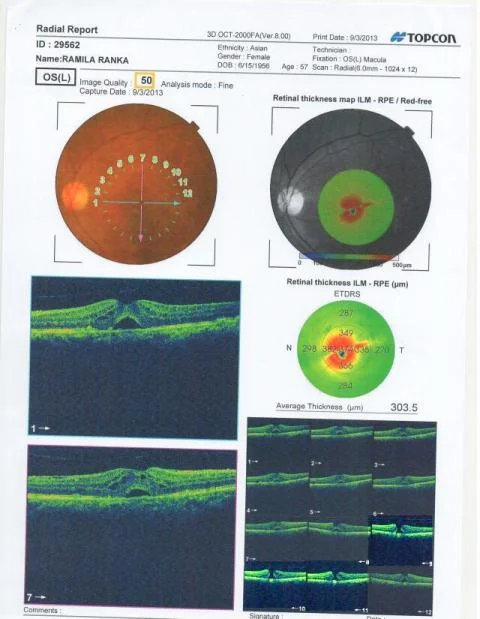
Home Role Of Optical Coherence Tomography This 57 years old female came to me with complaint of left eye diminished and wavy vision since 20 days. On examination her left eye retina showed drusen, fluid? Haemorrhage. Previous OCT done elsewhere showed Cystic lesion at macula. OCT repeated at our clinic showed Full Thickness Macular Hole. If one refers to the OCT done elsewhere, scan 6, 7, 8 shows Macular Hole.
Treatment Of Diabetic Macular Edema Without Intravitreal Injections
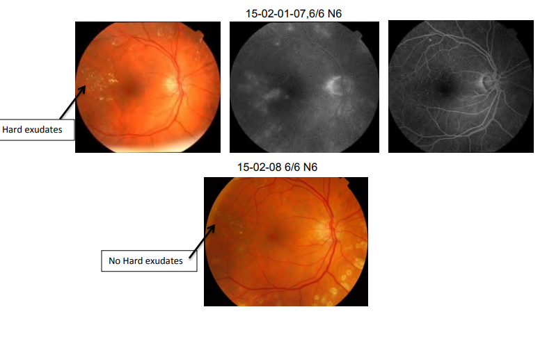
Home Treatment Of Diabetic Macular Edema Without Intravitreal Injections Click here to open PDF
Uses Of Fundus Autofluorescence
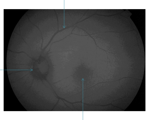
Home Uses Of Fundus Autofluorescence Click here to open PDF
Whether Gauge Is Important In Vitreous Surgery ?
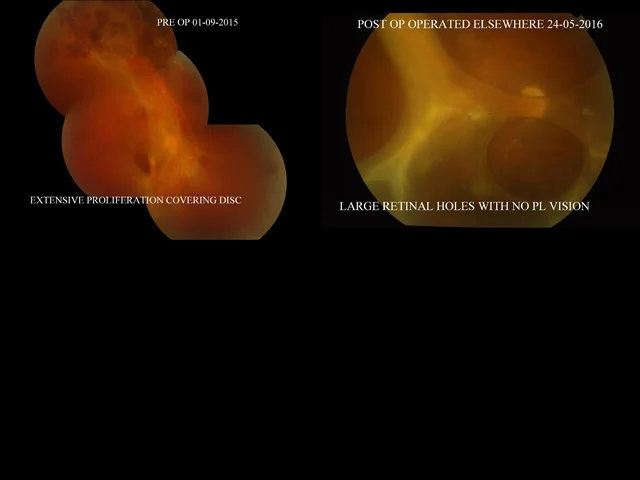
Home Whether Gauge Is Important In Vitreous Surgery ? I would like to share a thought with you today the trend is towards 27g vitrectomy with 10,000 to 12,000 cut rate, which is probably also commercially driven to remain ahead in the race. But the simple fact remains that a good surgery is a must irrespective of 23, 25 or 27gauge vitrectomy. The first patient 20years old seen by us a year back had advanced fibro vascular membrane over disc & going superiorly. He underwent vitrectomy elsewhere with 27g & has large holes in retina & No PL vision in the eye post-surgery. The second patient 21 years old had advanced fibro vascular membrane from the disc going superiorly with tractional detachment. He underwent 23g vitrectomy at our place & has recovered with 6/36 N8p vision post op. Both eyes patient had Eale’s Disease
Good Effect Of Inj. Eylea In Pigment Epithelial Detachment Due To Pcv In Wet Age Related Macular Degeneration
Home Spontaneous Closure Of Macular Hole Despite Presence Of Posterior Vitreous Detachment Pre Macular Hole Formation This 73 Yrs old lady is One Eyed and has Age Related Macular Degeneration with RPE Detachment. She was treated earlier with Inj. Accentrix & Inj. Avastin. She underwent Eylea injection following which she has responded well and right now the disease under control.
Spontaneous Closure Of Macular Hole Despite Presence Of Posterior Vitreous Detachment Pre Macular Hole Formation
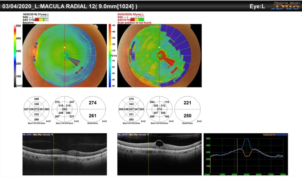
Home Spontaneous Closure Of Macular Hole Despite Presence Of Posterior Vitreous Detachment Pre Macular Hole Formation I would like to share this unusual picture of 52 yr old myopic (-5.0 ds) , Vision in left eye in 2019 was 6/6 N6 Optical coherence tomography reveled normal foveal anatomy with posterior vitreoud detachment, On April 2020 patient became symptomatic for vision probleme in left eye Left eye vision dropped to 6/12 N10 , Optical coherence tomography showed macular cyst with inner hole , complete posterior vitreoud detachment, On July 2020 vision in left eye 6/9 N6 Optical coherence tomography almost normal Patient symptomaticsally better Treatment given only Nepafenac eye drops Other eye normal It is spontaneous resolution of early macular hole but surprisingly there is posterior vitreoud detachment, since a year